- PDB-9mov: Cryo-EM structure of factor Va bound to activated protein C -
+
データを開く
IDまたはキーワード:
読み込み中...
-
基本情報
登録情報
データベース: PDB / ID: 9mov
タイトル
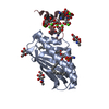
Cryo-EM structure of factor Va bound to activated protein C
要素
(Coagulation factor Va ...) x 2
(Vitamin K-dependent protein ...) x 2
キーワード
BLOOD CLOTTING / Coagulation / Activated Factor V / Activated Protein C
機能・相同性
機能・相同性情報
activated protein C (thrombin-activated peptidase) / positive regulation of establishment of endothelial barrier / negative regulation of coagulation / response to vitamin K / platelet alpha granule / Cargo concentration in the ER / COPII-coated ER to Golgi transport vesicle / COPII-mediated vesicle transport / blood circulation / negative regulation of blood coagulation ...activated protein C (thrombin-activated peptidase) / positive regulation of establishment of endothelial barrier / negative regulation of coagulation / response to vitamin K / platelet alpha granule / Cargo concentration in the ER / COPII-coated ER to Golgi transport vesicle / COPII-mediated vesicle transport / blood circulation / negative regulation of blood coagulation / Transport of gamma-carboxylated protein precursors from the endoplasmic reticulum to the Golgi apparatus / : / Gamma-carboxylation of protein precursors / Removal of aminoterminal propeptides from gamma-carboxylated proteins / : / endoplasmic reticulum-Golgi intermediate compartment membrane / platelet alpha granule lumen / Cell surface interactions at the vascular wall / Post-translational protein phosphorylation / Golgi lumen / negative regulation of inflammatory response / Regulation of Insulin-like Growth Factor (IGF) transport and uptake by Insulin-like Growth Factor Binding Proteins (IGFBPs) / blood coagulation / Platelet degranulation / extracellular vesicle / endoplasmic reticulum lumen / copper ion binding / serine-type endopeptidase activity / calcium ion binding / negative regulation of apoptotic process / endoplasmic reticulum / Golgi apparatus / proteolysis / : / extracellular region / membrane / plasma membrane 類似検索 - 分子機能
Coagulation factor 5/8-like / : / Coagulation factors 5/8 type C domain (FA58C) signature 2. / Multicopper oxidases, conserved site / Multicopper oxidases signature 1. / Coagulation factors 5/8 type C domain (FA58C) signature 1. / Coagulation factor 5/8 C-terminal domain, discoidin domain / Coagulation factors 5/8 type C domain (FA58C) profile. / Peptidase S1A, coagulation factor VII/IX/X/C/Z / F5/8 type C domain ...Coagulation factor 5/8-like / : / Coagulation factors 5/8 type C domain (FA58C) signature 2. / Multicopper oxidases, conserved site / Multicopper oxidases signature 1. / Coagulation factors 5/8 type C domain (FA58C) signature 1. / Coagulation factor 5/8 C-terminal domain, discoidin domain / Coagulation factors 5/8 type C domain (FA58C) profile. / Peptidase S1A, coagulation factor VII/IX/X/C/Z / F5/8 type C domain / : / Coagulation factor 5/8 C-terminal domain / Coagulation factor-like, Gla domain superfamily / Multicopper oxidase, N-terminal / Multicopper oxidase / Coagulation Factor Xa inhibitory site / EGF-type aspartate/asparagine hydroxylation site / EGF-like calcium-binding, conserved site / Calcium-binding EGF-like domain signature. / Aspartic acid and asparagine hydroxylation site. / EGF-like calcium-binding domain / Calcium-binding EGF-like domain / Vitamin K-dependent carboxylation/gamma-carboxyglutamic (GLA) domain / Gamma-carboxyglutamic acid-rich (GLA) domain / Gamma-carboxyglutamic acid-rich (GLA) domain superfamily / Vitamin K-dependent carboxylation domain. / Gla domain profile. / Domain containing Gla (gamma-carboxyglutamate) residues. / Epidermal growth factor-like domain. / EGF-like domain profile. / EGF-like domain signature 1. / Cupredoxin / EGF-like domain signature 2. / EGF-like domain / Galactose-binding-like domain superfamily / Serine proteases, trypsin family, histidine active site / Serine proteases, trypsin family, serine active site / Serine proteases, trypsin family, histidine active site. / Peptidase S1A, chymotrypsin family / Serine proteases, trypsin family, serine active site. / Serine proteases, trypsin domain profile. / Trypsin-like serine protease / Serine proteases, trypsin domain / Trypsin / Peptidase S1, PA clan, chymotrypsin-like fold / Peptidase S1, PA clan 類似検索 - ドメイン・相同性
Vitamin K-dependent protein C / Coagulation factor V 類似検索 - 構成要素
National Institutes of Health/National Heart, Lung, and Blood Institute (NIH/NHLBI)
HL049413, HL139554 and HL147821
米国
Childrens Discovery Institute of Washington University and St. Louis Childrens Hospital
CDI-CORE-2015-505 and CDI-CORE-2019-813
米国
The Foundation for Barnes-Jewish Hospital
3770
米国
National Institutes of Health/National Institute of Diabetes and Digestive and Kidney Disease (NIH/NIDDK)
DK020579
米国
National Institutes of Health/National Cancer Institute (NIH/NCI)
CA091842
米国
引用
ジャーナル: Blood / 年: 2025 タイトル: Cryo-EM structure of coagulation factor Va bound to activated protein C. 著者: Bassem M Mohammed / Katherine Basore / Enrico Di Cera / 要旨: Coagulation factor Va (FVa) is the cofactor component of the prothrombinase complex required for rapid generation of thrombin from prothrombin in the penultimate step of the coagulation cascade. In ...Coagulation factor Va (FVa) is the cofactor component of the prothrombinase complex required for rapid generation of thrombin from prothrombin in the penultimate step of the coagulation cascade. In addition, FVa is a target for proteolytic inactivation by activated protein C (APC). Like other protein-protein interactions in the coagulation cascade, the FVa-APC interaction has long posed a challenge to structural biology and its molecular underpinnings remain unknown. A recent cryogenic electron microscopy (cryo-EM) structure of FVa has revealed the arrangement of its A1-A2-A3-C1-C2 domains and the environment of the sites of APC cleavage at R306 and R506. Here, we report the cryo-EM structure of the FVa-APC complex at 3.15 Å resolution in which the protease domain of APC engages R506 in the A2 domain of FVa through electrostatic interactions between positively charged residues in the 30-loop and 70-loop of APC and an electronegative surface of FVa. The auxiliary γ-carboxyglutamic acid and epidermal growth factor domains of APC are highly dynamic and point to solvent, without making contacts with FVa. Binding of APC displaces a large portion of the A2 domain of FVa and projects the 654VKCIPDDDEDSYEIFEP670 segment as a "latch," or exosite ligand, over the 70-loop of the enzyme. The latch induces a large conformational change of the autolysis loop of APC, which in turn promotes docking of R506 into the primary specificity pocket. The cryo-EM structure of the FVa-APC complex validates the bulk of existing biochemical data and offers molecular context for a key regulatory interaction of the coagulation cascade.
A: Coagulation factor Va heavy chain B: Coagulation factor Va light chain C: Vitamin K-dependent protein C D: Vitamin K-dependent protein C heavy chain ヘテロ分子
 ムービー
ムービー コントローラー


 データを開く
 基本情報
基本情報 要素
要素 キーワード
キーワード 機能・相同性情報
 Homo sapiens (ヒト)
 データ登録者
 米国, 5件
  引用
引用 構造の表示
構造の表示 ダウンロードとリンク
 その他のダウンロード


 PDBj
PDBj


 集合体
集合体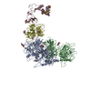
 Mesocricetus auratus (ネズミ)

 タイプ: D-saccharide, beta linking / 分子量: 221.208 Da / 分子数: 9 / 由来タイプ: 天然 / 式: C8H15NO6 / タイプ: SUBJECT OF INVESTIGATION
 分子量: 40.078 Da / 分子数: 7 / 由来タイプ: 合成 / 式: Ca / タイプ: SUBJECT OF INVESTIGATION
 試料調製
試料調製 電子顕微鏡撮影
 FIELD EMISSION GUN / 加速電圧: 300 kV / 照射モード: FLOOD BEAM
 解析
解析