Movie
Movie Controller

[English] 日本語
 Yorodumi
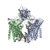
- PDB-9i5k: Structure of the Chaetomium thermophilum Pmt4 homodimer (C2 symmetry) -
+ Open data

- Basic information
| Entry | Database: PDB / ID: 9i5k | ||||||||||||||||||||||||||||||
|---|---|---|---|---|---|---|---|---|---|---|---|---|---|---|---|---|---|---|---|---|---|---|---|---|---|---|---|---|---|---|---|
| Title | Structure of the Chaetomium thermophilum Pmt4 homodimer (C2 symmetry) | ||||||||||||||||||||||||||||||
 Components Components | Dolichyl-phosphate-mannose--protein mannosyltransferase | ||||||||||||||||||||||||||||||
 Keywords Keywords | MEMBRANE PROTEIN / homodimer / glycosylation / ER / biogenesis | ||||||||||||||||||||||||||||||
| Function / homology |  Function and homology information Function and homology informationdolichyl-phosphate-mannose-protein mannosyltransferase / dolichyl-phosphate-mannose-protein mannosyltransferase activity / endoplasmic reticulum membrane Similarity search - Function | ||||||||||||||||||||||||||||||
| Biological species |  Thermochaetoides thermophila (fungus) Thermochaetoides thermophila (fungus) | ||||||||||||||||||||||||||||||
| Method | ELECTRON MICROSCOPY / single particle reconstruction / cryo EM / Resolution: 3.2 Å | ||||||||||||||||||||||||||||||
 Authors Authors | McDowell, M.A. / Wild, K. / Sinning, I. | ||||||||||||||||||||||||||||||
| Funding support |  Germany, 1items Germany, 1items
| ||||||||||||||||||||||||||||||
 Citation Citation | Journal: Nat Commun / Year: 2025 Title: Structural characterisation of the fungal Pmt4 homodimer. Authors: Melanie A McDowell / Klemens Wild / Francesco Fiorentino / Daniela Bausewein / Anke Metschies / Antonella Chiapparino / Yvonne Hackmann / Florestan L Bilsing / David Brenske / Sofia ...Authors: Melanie A McDowell / Klemens Wild / Francesco Fiorentino / Daniela Bausewein / Anke Metschies / Antonella Chiapparino / Yvonne Hackmann / Florestan L Bilsing / David Brenske / Sofia Mortensen / Di Wu / Carol V Robinson / Sabine Strahl / Irmgard Sinning /   Abstract: Protein O-mannosyltransferases (PMTs) are conserved endoplasmic reticulum membrane-embedded enzymes responsible for the transfer of mannose from dolichol phosphate-mannose (Dol-P-Man) to ...Protein O-mannosyltransferases (PMTs) are conserved endoplasmic reticulum membrane-embedded enzymes responsible for the transfer of mannose from dolichol phosphate-mannose (Dol-P-Man) to serine/threonine-rich protein substrates or unfolded proteins. PMTs from three subfamilies form obligate dimers with different substrate specificities and require the concerted action of their transmembrane domains (TMDs) and a luminal MIR domain for catalysis. Here, we present structures, native mass spectrometry, and structure-based mutagenesis of the fungal Pmt4 homodimer. The core fold of the TMDs and MIR domain is conserved with the Pmt1-Pmt2 heterodimer, indicating a shared catalytic mechanism. Distinct from Pmt4, the MIR domain interacts in cis with the TMDs of the same subunit and has a β-hairpin insertion required for O-mannosylation of substrates. We further identify a cytosolic binding site for substrate Dol-P-Man within the Pmt4 TMDs, which is conserved amongst PMTs and important for in vivo activity. Thus, we provide a framework to understand the substrate specificity and regulation of the Pmt4 homodimer. | ||||||||||||||||||||||||||||||
| History |
|
- Structure visualization
| Structure viewer | Molecule: MolmilJmol/JSmol |
|---|
- Downloads & links
-Download
| PDBx/mmCIF format | 9i5k.cif.gz | 202.2 KB | Display | PDBx/mmCIF format |
|---|---|---|---|---|
| PDB format | pdb9i5k.ent.gz | Display | PDB format | |
| PDBx/mmJSON format | 9i5k.json.gz | Tree view | PDBx/mmJSON format | |
| Others |  Other downloads Other downloads |
-Validation report
| Arichive directory | https://data.pdbj.org/pub/pdb/validation_reports/i5/9i5kftp://data.pdbj.org/pub/pdb/validation_reports/i5/9i5k | HTTPS FTP |
|---|
-Related structure data
| Related structure data |  52631MC  9fd0C  9fd1C  9i5lC C: citing same article ( M: map data used to model this data |
|---|---|
| Similar structure data |

-Links
 PDBj
PDBj- Assembly
| Deposited unit | 
|
|---|---|
| 1 |
|
-Components
| #1: Protein | Mass: 92046.484 Da / Num. of mol.: 2 Source method: isolated from a genetically manipulated source Source: (gene. exp.) Thermochaetoides thermophila (fungus) / Gene: CTHT_0059600 / Production host:  References: UniProt: G0SET1, dolichyl-phosphate-mannose-protein mannosyltransferase #2: Chemical | Mass: 684.880 Da / Num. of mol.: 2 / Source method: obtained synthetically / Formula: C37H65O9P #3: Chemical | Mass: 658.974 Da / Num. of mol.: 2 / Source method: obtained synthetically / Formula: C41H71O4P Has ligand of interest | Y | Has protein modification | N | |
|---|
-Experimental details
-Experiment
| Experiment | Method: ELECTRON MICROSCOPY |
|---|---|
| EM experiment | Aggregation state: PARTICLE / 3D reconstruction method: single particle reconstruction |
- Sample preparation
| Component | Name: Chaetomium thermophilum Pmt4 homodimer bound to dolichol-phosphate and dolichol-phosphate-mannose Type: COMPLEX / Entity ID: #1 / Source: RECOMBINANT |
|---|---|
| Molecular weight | Value: 0.18 MDa / Experimental value: NO |
| Source (natural) | Organism: Thermochaetoides thermophila (fungus) |
| Source (recombinant) | Organism: |
| Buffer solution | pH: 7.5 Details: 20 mM HEPES pH 7.5 200 mM NaCl 0.01% (w/v) LMNG 0.001% (w/v) cholesterol |
| Specimen | Conc.: 1.7 mg/ml / Embedding applied: NO / Shadowing applied: NO / Staining applied: NO / Vitrification applied: YES |
| Specimen support | Grid material: COPPER / Grid mesh size: 300 divisions/in. / Grid type: Quantifoil R1.2/1.3 |
| Vitrification | Instrument: FEI VITROBOT MARK IV / Cryogen name: ETHANE / Humidity: 100 % / Chamber temperature: 279 K |
- Electron microscopy imaging
| Experimental equipment |  Model: Titan Krios / Image courtesy: FEI Company |
|---|---|
| Microscopy | Model: TFS KRIOS |
| Electron gun | Electron source:  FIELD EMISSION GUN / Accelerating voltage: 300 kV / Illumination mode: FLOOD BEAM FIELD EMISSION GUN / Accelerating voltage: 300 kV / Illumination mode: FLOOD BEAM |
| Electron lens | Mode: BRIGHT FIELD / Nominal defocus max: 2000 nm / Nominal defocus min: 800 nm |
| Image recording | Electron dose: 42.7 e/Å2 / Film or detector model: GATAN K2 SUMMIT (4k x 4k) / Num. of grids imaged: 1 / Num. of real images: 9879 |
| Image scans | Movie frames/image: 40 |
- Processing
| EM software |
| ||||||||||||||||||||||||||||||||||||||||
|---|---|---|---|---|---|---|---|---|---|---|---|---|---|---|---|---|---|---|---|---|---|---|---|---|---|---|---|---|---|---|---|---|---|---|---|---|---|---|---|---|---|
| CTF correction | Type: PHASE FLIPPING AND AMPLITUDE CORRECTION | ||||||||||||||||||||||||||||||||||||||||
| Particle selection | Num. of particles selected: 2199173 | ||||||||||||||||||||||||||||||||||||||||
| Symmetry | Point symmetry: C2 (2 fold cyclic) | ||||||||||||||||||||||||||||||||||||||||
| 3D reconstruction | Resolution: 3.2 Å / Resolution method: FSC 0.143 CUT-OFF / Num. of particles: 210070 / Symmetry type: POINT | ||||||||||||||||||||||||||||||||||||||||
| Atomic model building | Space: REAL | ||||||||||||||||||||||||||||||||||||||||
| Atomic model building | PDB-ID: 6P25 Pdb chain-ID: B / Accession code: 6P25 / Details: SCWRL homology model / Source name: PDB / Type: experimental model | ||||||||||||||||||||||||||||||||||||||||
| Refine LS restraints |
|