 Movie
Movie Controller

[English] 日本語
 Yorodumi
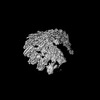
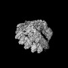
- PDB-9g40: Structure of the Position 7 CMG-decorated gamma-Tubulin Ring Comp... -
+ Open data

- Basic information
| Entry | Database: PDB / ID: 9g40 | |||||||||
|---|---|---|---|---|---|---|---|---|---|---|
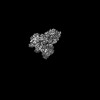
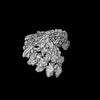
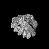
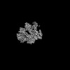
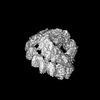
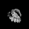
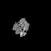
| Title | Structure of the Position 7 CMG-decorated gamma-Tubulin Ring Complex from Pig Brain | |||||||||
 Components Components |
| |||||||||
 Keywords Keywords | STRUCTURAL PROTEIN / Tubulin Complex | |||||||||
| Function / homology |  Function and homology information Function and homology informationRecruitment of mitotic centrosome proteins and complexes / microtubule organizing center organization / negative regulation of centriole replication / equatorial microtubule organizing center / regulation of mitotic cell cycle spindle assembly checkpoint / microtubule plus-end / polar microtubule / gamma-tubulin complex / microtubule nucleation / microtubule bundle formation ...Recruitment of mitotic centrosome proteins and complexes / microtubule organizing center organization / negative regulation of centriole replication / equatorial microtubule organizing center / regulation of mitotic cell cycle spindle assembly checkpoint / microtubule plus-end / polar microtubule / gamma-tubulin complex / microtubule nucleation / microtubule bundle formation / gamma-tubulin binding / centrosome cycle / Recruitment of NuMA to mitotic centrosomes / regulation of neuron differentiation / pericentriolar material / mitotic spindle pole / establishment of mitotic spindle orientation / centriole replication / negative regulation of neuron differentiation / spindle assembly / cytoplasmic microtubule organization / Loss of Nlp from mitotic centrosomes / Loss of proteins required for interphase microtubule organization from the centrosome / Recruitment of mitotic centrosome proteins and complexes / Recruitment of NuMA to mitotic centrosomes / Anchoring of the basal body to the plasma membrane / positive regulation of microtubule polymerization / centriole / AURKA Activation by TPX2 / tubulin binding / neurogenesis / meiotic cell cycle / chromosome segregation / neuron migration / brain development / spindle / spindle pole / microtubule cytoskeleton organization / Regulation of PLK1 Activity at G2/M Transition / cell junction / mitotic cell cycle / microtubule binding / microtubule / cytoskeleton / transcription cis-regulatory region binding / calmodulin binding / centrosome / protein-containing complex binding / protein kinase binding / perinuclear region of cytoplasm / positive regulation of DNA-templated transcription / Golgi apparatus / extracellular exosome / nucleoplasm / cytosol / cytoplasm Similarity search - Function | |||||||||
| Biological species |  Homo sapiens (human) Homo sapiens (human) | |||||||||
| Method | ELECTRON MICROSCOPY / single particle reconstruction / cryo EM / Resolution: 4.3 Å | |||||||||
 Authors Authors | Munoz-Hernandez, H. / Krutyholowa, R. / Wieczorek, M. | |||||||||
| Funding support |  Switzerland, 2items Switzerland, 2items
| |||||||||
 Citation Citation | Journal: Dev Cell / Year: 2024 Title: Partial closure of the γ-tubulin ring complex by CDK5RAP2 activates microtubule nucleation. Authors: Yixin Xu / Hugo Muñoz-Hernández / Rościsław Krutyhołowa / Florina Marxer / Ferdane Cetin / Michal Wieczorek / Abstract: Microtubule nucleation is templated by the γ-tubulin ring complex (γ-TuRC), but its structure deviates from the geometry of α-/β-tubulin in the microtubule, explaining the complex's poor ...Microtubule nucleation is templated by the γ-tubulin ring complex (γ-TuRC), but its structure deviates from the geometry of α-/β-tubulin in the microtubule, explaining the complex's poor nucleating activity. Several proteins may activate the γ-TuRC, but the mechanisms underlying activation are not known. Here, we determined the structure of the porcine γ-TuRC purified using CDK5RAP2's centrosomin motif 1 (CM1). We identified an unexpected conformation of the γ-TuRC bound to multiple protein modules containing MZT2, GCP2, and CDK5RAP2, resulting in a long-range constriction of the γ-tubulin ring that brings it in closer agreement with the 13-protofilament microtubule. Additional CDK5RAP2 promoted γ-TuRC decoration and stimulated the microtubule-nucleating activities of the porcine γ-TuRC and a reconstituted, CM1-free human complex in single-molecule assays. Our results provide a structural mechanism for the control of microtubule nucleation by CM1 proteins and identify conformational transitions in the γ-TuRC that prime it for microtubule nucleation. | |||||||||
| History |
|
- Structure visualization
| Structure viewer | Molecule: MolmilJmol/JSmol |
|---|
- Downloads & links
-Download
| PDBx/mmCIF format | 9g40.cif.gz | 593.1 KB | Display | PDBx/mmCIF format |
|---|---|---|---|---|
| PDB format | pdb9g40.ent.gz | 446 KB | Display | PDB format |
| PDBx/mmJSON format | 9g40.json.gz | Tree view | PDBx/mmJSON format | |
| Others |  Other downloads Other downloads |
-Validation report
| Summary document | 9g40_validation.pdf.gz | 1.3 MB | Display | wwPDB validaton report |
|---|---|---|---|---|
| Full document | 9g40_full_validation.pdf.gz | 1.4 MB | Display | |
| Data in XML | 9g40_validation.xml.gz | 52.2 KB | Display | |
| Data in CIF | 9g40_validation.cif.gz | 76.5 KB | Display | |
| Arichive directory | https://data.pdbj.org/pub/pdb/validation_reports/g4/9g40ftp://data.pdbj.org/pub/pdb/validation_reports/g4/9g40 | HTTPS FTP |
-Related structure data
| Related structure data | 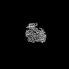 51020MC 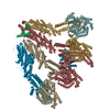 9g3xC 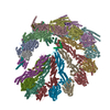 9g3yC 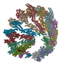 9g3zC M: map data used to model this data C: citing same article ( |
|---|---|
| Similar structure data |


-Links
 PDBj
PDBj- Assembly
| Deposited unit | 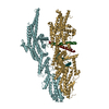
|
|---|---|
| 1 |
|
-Components
| #1: Protein | Mass: 103172.477 Da / Num. of mol.: 1 / Source method: isolated from a natural source / Details: SsGCP3 / Source: (natural) | ||
|---|---|---|---|
| #2: Protein | Mass: 102609.703 Da / Num. of mol.: 1 / Source method: isolated from a natural source / Source: (natural) | ||
| #3: Protein | Mass: 15920.321 Da / Num. of mol.: 1 / Source method: isolated from a natural source / Source: (natural) | ||
| #4: Protein | Mass: 215344.219 Da / Num. of mol.: 2 Source method: isolated from a genetically manipulated source Source: (gene. exp.) Homo sapiens (human) / Gene: CDK5RAP2, CEP215, KIAA1633 / Details (production host): (Kan) / Production host:  Has protein modification | N | |
-Experimental details
-Experiment
| Experiment | Method: ELECTRON MICROSCOPY |
|---|---|
| EM experiment | Aggregation state: PARTICLE / 3D reconstruction method: single particle reconstruction |
- Sample preparation
| Component | Name: Gamma-Tubulin Ring Complex in native pig brain / Type: COMPLEX / Entity ID: all / Source: NATURAL | ||||||||||||||||||||||||||||||||
|---|---|---|---|---|---|---|---|---|---|---|---|---|---|---|---|---|---|---|---|---|---|---|---|---|---|---|---|---|---|---|---|---|---|
| Molecular weight | Experimental value: NO | ||||||||||||||||||||||||||||||||
| Source (natural) | Organism: | ||||||||||||||||||||||||||||||||
| Buffer solution | pH: 7.5 Details: Listed in "g-TuRC cryo-EM sample preparation" section. | ||||||||||||||||||||||||||||||||
| Buffer component |
| ||||||||||||||||||||||||||||||||
| Specimen | Embedding applied: NO / Shadowing applied: NO / Staining applied: NO / Vitrification applied: YES | ||||||||||||||||||||||||||||||||
| Vitrification | Instrument: FEI VITROBOT MARK III / Cryogen name: ETHANE-PROPANE / Humidity: 100 % / Chamber temperature: 277.15 K |
- Electron microscopy imaging
| Experimental equipment |  Model: Titan Krios / Image courtesy: FEI Company |
|---|---|
| Microscopy | Model: TFS KRIOS |
| Electron gun | Electron source:  FIELD EMISSION GUN / Accelerating voltage: 300 kV / Illumination mode: SPOT SCAN FIELD EMISSION GUN / Accelerating voltage: 300 kV / Illumination mode: SPOT SCAN |
| Electron lens | Mode: BRIGHT FIELD / Nominal defocus max: 2700 nm / Nominal defocus min: 900 nm |
| Image recording | Electron dose: 55 e/Å2 / Film or detector model: GATAN K3 BIOQUANTUM (6k x 4k) |
- Processing
| CTF correction | Type: NONE | ||||||||||||||||||||||||
|---|---|---|---|---|---|---|---|---|---|---|---|---|---|---|---|---|---|---|---|---|---|---|---|---|---|
| 3D reconstruction | Resolution: 4.3 Å / Resolution method: FSC 0.143 CUT-OFF / Num. of particles: 93207 / Symmetry type: POINT | ||||||||||||||||||||||||
| Refine LS restraints |
|
