 Movie
Movie Controller

[English] 日本語
 Yorodumi
- PDB-9f5z: Structure of the Chlamydomonas reinhardtii respiratory complex II... -
+ Open data

- Basic information
| Entry | Database: PDB / ID: 9f5z | |||||||||||||||||||||||||||
|---|---|---|---|---|---|---|---|---|---|---|---|---|---|---|---|---|---|---|---|---|---|---|---|---|---|---|---|---|
| Title | Structure of the Chlamydomonas reinhardtii respiratory complex III from respiratory supercomplex | |||||||||||||||||||||||||||
 Components Components |
| |||||||||||||||||||||||||||
 Keywords Keywords | MEMBRANE PROTEIN / Mitochondria / respiratory complex / respiration / alga | |||||||||||||||||||||||||||
| Function / homology |  Function and homology information Function and homology informationquinol-cytochrome-c reductase / mitochondrial processing peptidase / respiratory chain complex III / quinol-cytochrome-c reductase / quinol-cytochrome-c reductase activity / mitochondrial electron transport, ubiquinol to cytochrome c / respiratory electron transport chain / metalloendopeptidase activity / 2 iron, 2 sulfur cluster binding / electron transfer activity ...quinol-cytochrome-c reductase / mitochondrial processing peptidase / respiratory chain complex III / quinol-cytochrome-c reductase / quinol-cytochrome-c reductase activity / mitochondrial electron transport, ubiquinol to cytochrome c / respiratory electron transport chain / metalloendopeptidase activity / 2 iron, 2 sulfur cluster binding / electron transfer activity / oxidoreductase activity / mitochondrial inner membrane / mitochondrial matrix / heme binding / mitochondrion / proteolysis / membrane / metal ion binding Similarity search - Function | |||||||||||||||||||||||||||
| Biological species |   Chlamydomonas reinhardtii (plant) Chlamydomonas reinhardtii (plant) | |||||||||||||||||||||||||||
| Method | ELECTRON MICROSCOPY / single particle reconstruction / cryo EM / Resolution: 2.39 Å | |||||||||||||||||||||||||||
 Authors Authors | Waltz, F. / Righetto, R. / Kotecha, A. / Engel, B.D. | |||||||||||||||||||||||||||
| Funding support |  Germany, Germany,  Switzerland, 2items Switzerland, 2items
| |||||||||||||||||||||||||||
 Citation Citation | Journal: Science / Year: 2025 Title: In-cell architecture of the mitochondrial respiratory chain. Authors: Florent Waltz / Ricardo D Righetto / Lorenz Lamm / Thalia Salinas-Giegé / Ron Kelley / Xianjun Zhang / Martin Obr / Sagar Khavnekar / Abhay Kotecha / Benjamin D Engel /    Abstract: Mitochondria regenerate adenosine triphosphate (ATP) through oxidative phosphorylation. This process is carried out by five membrane-bound complexes collectively known as the respiratory chain, ...Mitochondria regenerate adenosine triphosphate (ATP) through oxidative phosphorylation. This process is carried out by five membrane-bound complexes collectively known as the respiratory chain, working in concert to transfer electrons and pump protons. The precise organization of these complexes in native cells is debated. We used in situ cryo-electron tomography to visualize the native structures and organization of several major mitochondrial complexes in cells. ATP synthases and respiratory complexes segregate into curved and flat crista membrane domains, respectively. Respiratory complexes I, III, and IV assemble into a respirasome supercomplex, from which we determined a native 5-angstrom (Å) resolution structure showing binding of electron carrier cytochrome . Combined with single-particle cryo-electron microscopy at 2.4-Å resolution, we model how the respiratory complexes organize inside native mitochondria. | |||||||||||||||||||||||||||
| History |
|
- Structure visualization
| Structure viewer | Molecule: MolmilJmol/JSmol |
|---|
- Downloads & links
-Download
| PDBx/mmCIF format | 9f5z.cif.gz | 857 KB | Display | PDBx/mmCIF format |
|---|---|---|---|---|
| PDB format | pdb9f5z.ent.gz | Display | PDB format | |
| PDBx/mmJSON format | 9f5z.json.gz | Tree view | PDBx/mmJSON format | |
| Others |  Other downloads Other downloads |
-Validation report
| Arichive directory | https://data.pdbj.org/pub/pdb/validation_reports/f5/9f5zftp://data.pdbj.org/pub/pdb/validation_reports/f5/9f5z | HTTPS FTP |
|---|
-Related structure data
| Related structure data |  50204MC  9f5xC  9f5yC 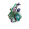 9f60C  9f61C  9f62C M: map data used to model this data C: citing same article ( |
|---|---|
| Similar structure data |


-Links
 PDBj
PDBj


- Assembly
| Deposited unit | 
|
|---|---|
| 1 |
|
-Components
-Protein , 5 types, 10 molecules 1A1B1E1F1G1H1M1N1Q1S
| #1: Protein | Mass: 42356.465 Da / Num. of mol.: 2 / Source method: isolated from a natural source / Source: (natural) Chlamydomonas reinhardtii (plant) / References: UniProt: P23662#3: Protein | Mass: 33868.195 Da / Num. of mol.: 2 / Source method: isolated from a natural source / Source: (natural) Chlamydomonas reinhardtii (plant) / References: UniProt: Q9FQ96, quinol-cytochrome-c reductase#4: Protein | Mass: 7040.211 Da / Num. of mol.: 2 / Source method: isolated from a natural source / Source: (natural) Chlamydomonas reinhardtii (plant) / References: UniProt: A8JC51#7: Protein | Mass: 55121.215 Da / Num. of mol.: 2 / Source method: isolated from a natural source / Source: (natural) Chlamydomonas reinhardtii (plant) / References: UniProt: A8J5P7#9: Protein | Mass: 49640.430 Da / Num. of mol.: 2 / Source method: isolated from a natural source / Source: (natural) Chlamydomonas reinhardtii (plant) / References: UniProt: A8IKI9 |
|---|
-Cytochrome b-c1 complex subunit ... , 3 types, 6 molecules 1C1D1I1J1R1T
| #2: Protein | Mass: 28545.494 Da / Num. of mol.: 2 / Source method: isolated from a natural source / Source: (natural) Chlamydomonas reinhardtii (plant) / References: UniProt: Q8HEB4, quinol-cytochrome-c reductase#5: Protein | Mass: 7991.032 Da / Num. of mol.: 2 / Source method: isolated from a natural source / Source: (natural) Chlamydomonas reinhardtii (plant) / References: UniProt: A8J8N9#10: Protein | Mass: 14062.162 Da / Num. of mol.: 2 / Source method: isolated from a natural source / Source: (natural) Chlamydomonas reinhardtii (plant) / References: UniProt: A8HX89 |
|---|
-Mitochondrial ubiquinol-cytochrome c oxidoreductase subunit ... , 2 types, 4 molecules 1K1L1O1P
| #6: Protein | Mass: 8678.994 Da / Num. of mol.: 2 / Source method: isolated from a natural source / Source: (natural) Chlamydomonas reinhardtii (plant) / References: UniProt: A8J7I9, quinol-cytochrome-c reductase#8: Protein | Mass: 6476.588 Da / Num. of mol.: 2 / Source method: isolated from a natural source / Source: (natural) Chlamydomonas reinhardtii (plant) / References: UniProt: A8J4K5, quinol-cytochrome-c reductase |
|---|
-Non-polymers , 8 types, 569 molecules 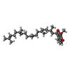


| #11: Chemical | ChemComp-UQ5 /  Mass: 522.758 Da / Num. of mol.: 4 / Source method: obtained synthetically / Formula: C34H50O4 Mass: 522.758 Da / Num. of mol.: 4 / Source method: obtained synthetically / Formula: C34H50O4#12: Chemical | ChemComp-HEC /  Mass: 618.503 Da / Num. of mol.: 6 / Source method: obtained synthetically / Formula: C34H34FeN4O4 Mass: 618.503 Da / Num. of mol.: 6 / Source method: obtained synthetically / Formula: C34H34FeN4O4#13: Chemical | ChemComp-CDL /  Mass: 1464.043 Da / Num. of mol.: 8 / Source method: obtained synthetically / Formula: C81H156O17P2 / Comment: phospholipid*YM Mass: 1464.043 Da / Num. of mol.: 8 / Source method: obtained synthetically / Formula: C81H156O17P2 / Comment: phospholipid*YM#14: Chemical | ChemComp-3PH /  Mass: 704.998 Da / Num. of mol.: 7 / Source method: obtained synthetically / Formula: C39H77O8P Mass: 704.998 Da / Num. of mol.: 7 / Source method: obtained synthetically / Formula: C39H77O8P#15: Chemical |  Mass: 734.039 Da / Num. of mol.: 2 / Source method: obtained synthetically / Formula: C40H80NO8P / Comment: phospholipid*YM Mass: 734.039 Da / Num. of mol.: 2 / Source method: obtained synthetically / Formula: C40H80NO8P / Comment: phospholipid*YM#16: Chemical |  Mass: 763.100 Da / Num. of mol.: 2 / Source method: obtained synthetically / Formula: C42H85NO8P / Comment: phospholipid*YM Mass: 763.100 Da / Num. of mol.: 2 / Source method: obtained synthetically / Formula: C42H85NO8P / Comment: phospholipid*YM#17: Chemical |  Mass: 65.409 Da / Num. of mol.: 2 / Source method: obtained synthetically / Formula: Zn Mass: 65.409 Da / Num. of mol.: 2 / Source method: obtained synthetically / Formula: Zn#18: Water | ChemComp-HOH / | Mass: 18.015 Da / Num. of mol.: 538 / Source method: isolated from a natural source / Formula: H2O |
|---|
-Details
| Has ligand of interest | N |
|---|---|
| Has protein modification | Y |
-Experimental details
-Experiment
| Experiment | Method: ELECTRON MICROSCOPY |
|---|---|
| EM experiment | Aggregation state: PARTICLE / 3D reconstruction method: single particle reconstruction |
- Sample preparation
| Component | Name: Chlamydomonas reinhardtii respirasome / Type: COMPLEX / Entity ID: #1-#10 / Source: NATURAL |
|---|---|
| Molecular weight | Experimental value: NO |
| Source (natural) | Organism: Chlamydomonas reinhardtii (plant) |
| Buffer solution | pH: 7.5 |
| Specimen | Conc.: 1 mg/ml / Embedding applied: NO / Shadowing applied: NO / Staining applied: NO / Vitrification applied: YES |
| Specimen support | Grid material: COPPER / Grid mesh size: 300 divisions/in. / Grid type: Quantifoil R2/1 |
| Vitrification | Instrument: FEI VITROBOT MARK IV / Cryogen name: ETHANE / Humidity: 100 % |
- Electron microscopy imaging
| Experimental equipment |  Model: Titan Krios / Image courtesy: FEI Company |
|---|---|
| Microscopy | Model: TFS KRIOS |
| Electron gun | Electron source:  FIELD EMISSION GUN / Accelerating voltage: 300 kV / Illumination mode: FLOOD BEAM FIELD EMISSION GUN / Accelerating voltage: 300 kV / Illumination mode: FLOOD BEAM |
| Electron lens | Mode: BRIGHT FIELD / Nominal defocus max: 2000 nm / Nominal defocus min: 500 nm |
| Image recording | Electron dose: 40 e/Å2 / Film or detector model: TFS FALCON 4i (4k x 4k) |
- Processing
| EM software |
| ||||||||||||||||||||||||||||||||
|---|---|---|---|---|---|---|---|---|---|---|---|---|---|---|---|---|---|---|---|---|---|---|---|---|---|---|---|---|---|---|---|---|---|
| CTF correction | Type: PHASE FLIPPING AND AMPLITUDE CORRECTION | ||||||||||||||||||||||||||||||||
| 3D reconstruction | Resolution: 2.39 Å / Resolution method: FSC 0.143 CUT-OFF / Num. of particles: 83443 / Symmetry type: POINT | ||||||||||||||||||||||||||||||||
| Refine LS restraints |
|
