 Movie
Movie Controller

[English] 日本語
 Yorodumi
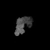
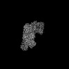
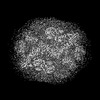
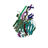
- EMDB-50211: Subtomogram average of the Chlamydomonas reinhardtii mitochondria... -
+ Open data

- Basic information
| Entry |  | |||||||||
|---|---|---|---|---|---|---|---|---|---|---|
| Title | Subtomogram average of the Chlamydomonas reinhardtii mitochondrial respirasome - C2 expanded | |||||||||
 Map data Map data | ||||||||||
 Sample Sample |
| |||||||||
 Keywords Keywords | respirasome / alga / respiration / mitochondria / MEMBRANE PROTEIN | |||||||||
| Biological species |   Chlamydomonas reinhardtii (plant) Chlamydomonas reinhardtii (plant) | |||||||||
| Method | subtomogram averaging / cryo EM / Resolution: 6.18 Å | |||||||||
 Authors Authors | Waltz F / Righetto R / Kotecha A / Engel BD | |||||||||
| Funding support |  Germany, Germany,  Switzerland, 2 items Switzerland, 2 items
| |||||||||
 Citation Citation | Journal: Science / Year: 2025 Title: In-cell architecture of the mitochondrial respiratory chain. Authors: Florent Waltz / Ricardo D Righetto / Lorenz Lamm / Thalia Salinas-Giegé / Ron Kelley / Xianjun Zhang / Martin Obr / Sagar Khavnekar / Abhay Kotecha / Benjamin D Engel /    Abstract: Mitochondria regenerate adenosine triphosphate (ATP) through oxidative phosphorylation. This process is carried out by five membrane-bound complexes collectively known as the respiratory chain, ...Mitochondria regenerate adenosine triphosphate (ATP) through oxidative phosphorylation. This process is carried out by five membrane-bound complexes collectively known as the respiratory chain, working in concert to transfer electrons and pump protons. The precise organization of these complexes in native cells is debated. We used in situ cryo-electron tomography to visualize the native structures and organization of several major mitochondrial complexes in cells. ATP synthases and respiratory complexes segregate into curved and flat crista membrane domains, respectively. Respiratory complexes I, III, and IV assemble into a respirasome supercomplex, from which we determined a native 5-angstrom (Å) resolution structure showing binding of electron carrier cytochrome . Combined with single-particle cryo-electron microscopy at 2.4-Å resolution, we model how the respiratory complexes organize inside native mitochondria. | |||||||||
| History |
|
- Structure visualization
| Supplemental images |
|---|

- Downloads & links
-EMDB archive
| Map data | emd_50211.map.gz | 8.9 MB |  EMDB map data format EMDB map data format | |
|---|---|---|---|---|
| Header (meta data) | emd-50211-v30.xmlemd-50211.xml | 17.5 KB 17.5 KB | Display Display | EMDB header |
| FSC (resolution estimation) | emd_50211_fsc.xml | 10.2 KB | Display | FSC data file |
| Images |  emd_50211.png emd_50211.png | 81.2 KB | ||
| Masks | emd_50211_msk_1.map | 91.1 MB | Mask map | |
| Filedesc metadata | emd-50211.cif.gz | 4.7 KB | ||
| Others | emd_50211_half_map_1.map.gzemd_50211_half_map_2.map.gz | 71.2 MB 71.2 MB | ||
| Archive directory |  http://ftp.pdbj.org/pub/emdb/structures/EMD-50211ftp://ftp.pdbj.org/pub/emdb/structures/EMD-50211 http://ftp.pdbj.org/pub/emdb/structures/EMD-50211ftp://ftp.pdbj.org/pub/emdb/structures/EMD-50211 | HTTPS FTP |
-Related structure data


-Links
| EMDB pages | EMDB (EBI/PDBe) / EMDataResource |
|---|
-Map
| File | Download / File: emd_50211.map.gz / Format: CCP4 / Size: 91.1 MB / Type: IMAGE STORED AS FLOATING POINT NUMBER (4 BYTES) | ||||||||||||||||||||||||||||||||||||
|---|---|---|---|---|---|---|---|---|---|---|---|---|---|---|---|---|---|---|---|---|---|---|---|---|---|---|---|---|---|---|---|---|---|---|---|---|---|
| Projections & slices | Image control
Images are generated by Spider. | ||||||||||||||||||||||||||||||||||||
| Voxel size | X=Y=Z: 1.91 Å | ||||||||||||||||||||||||||||||||||||
| Density |
| ||||||||||||||||||||||||||||||||||||
| Symmetry | Space group: 1 | ||||||||||||||||||||||||||||||||||||
| Details | EMDB XML:
|
 Z (Sec.)
 Y (Row.)
 X (Col.)


-Supplemental data
-Mask #1
| File | emd_50211_msk_1.map | ||||||||||||
|---|---|---|---|---|---|---|---|---|---|---|---|---|---|
| Projections & Slices |
| ||||||||||||
| Density Histograms |


-Half map: #1
| File | emd_50211_half_map_1.map | ||||||||||||
|---|---|---|---|---|---|---|---|---|---|---|---|---|---|
| Projections & Slices |
| ||||||||||||
| Density Histograms |


-Half map: #2
| File | emd_50211_half_map_2.map | ||||||||||||
|---|---|---|---|---|---|---|---|---|---|---|---|---|---|
| Projections & Slices |
| ||||||||||||
| Density Histograms |


- Sample components
-Entire : Chlamydomonas reinhardtii
| Entire | Name: Chlamydomonas reinhardtii (plant) |
|---|---|
| Components |
|
-Supramolecule #1: Chlamydomonas reinhardtii
| Supramolecule | Name: Chlamydomonas reinhardtii / type: cell / ID: 1 / Parent: 0 |
|---|---|
| Source (natural) | Organism: Chlamydomonas reinhardtii (plant) / Strain: CC-3994 mat3-4 mt+ |
-Experimental details
-Structure determination
| Method | cryo EM |
|---|---|
 Processing Processing | subtomogram averaging |
| Aggregation state | cell |
-Sample preparation
| Buffer | pH: 7 |
|---|---|
| Grid | Model: Quantifoil R2/1 / Material: COPPER / Support film - Material: CARBON / Pretreatment - Type: GLOW DISCHARGE / Pretreatment - Time: 20 sec. / Pretreatment - Atmosphere: AIR |
| Vitrification | Cryogen name: ETHANE-PROPANE / Chamber humidity: 100 % / Instrument: FEI VITROBOT MARK IV |
- Electron microscopy
| Microscope | TFS KRIOS |
|---|---|
| Specialist optics | Energy filter - Name: TFS Selectris X |
| Image recording | Film or detector model: TFS FALCON 4i (4k x 4k) / Digitization - Dimensions - Width: 4096 pixel / Digitization - Dimensions - Height: 4096 pixel / Average electron dose: 3.5 e/Å2 |
| Electron beam | Acceleration voltage: 300 kV / Electron source:  FIELD EMISSION GUN FIELD EMISSION GUN |
| Electron optics | Illumination mode: FLOOD BEAM / Imaging mode: BRIGHT FIELD / Nominal defocus max: 3.5 µm / Nominal defocus min: 0.8 µm / Nominal magnification: 64000 |
| Sample stage | Specimen holder model: FEI TITAN KRIOS AUTOGRID HOLDER / Cooling holder cryogen: NITROGEN |
| Experimental equipment |  Model: Titan Krios / Image courtesy: FEI Company |

