 Movie
Movie Controller

[English] 日本語
 Yorodumi
- PDB-8u72: Cryo-EM structure of the SPARTA oligomer with guide RNA and target DNA -
+ Open data

- Basic information
| Entry | Database: PDB / ID: 8u72 | |||||||||||||||||||||
|---|---|---|---|---|---|---|---|---|---|---|---|---|---|---|---|---|---|---|---|---|---|---|
| Title | Cryo-EM structure of the SPARTA oligomer with guide RNA and target DNA | |||||||||||||||||||||
 Components Components |
| |||||||||||||||||||||
 Keywords Keywords | RNA BINDING PROTEIN/RNA/DNA / RNA / DNA / Argonaute / TIR / immune system / RNA BINDING PROTEIN-RNA-DNA complex | |||||||||||||||||||||
| Function / homology |  Function and homology information Function and homology information | |||||||||||||||||||||
| Biological species |  Thermoflavifilum thermophilum (bacteria) Thermoflavifilum thermophilum (bacteria)synthetic construct (others) | |||||||||||||||||||||
| Method | ELECTRON MICROSCOPY / single particle reconstruction / cryo EM / Resolution: 3.15 Å | |||||||||||||||||||||
 Authors Authors | Malik, R. / Kottur, J. / Aggarwal, A.K. | |||||||||||||||||||||
| Funding support |  United States, 1items United States, 1items
| |||||||||||||||||||||
 Citation Citation | Journal: Nat Commun / Year: 2024 Title: Nucleic acid mediated activation of a short prokaryotic Argonaute immune system. Authors: Jithesh Kottur / Radhika Malik / Aneel K Aggarwal /  Abstract: A short prokaryotic Argonaute (pAgo) TIR-APAZ (SPARTA) defense system, activated by invading DNA to unleash its TIR domain for NAD(P) hydrolysis, was recently identified in bacteria. We report the ...A short prokaryotic Argonaute (pAgo) TIR-APAZ (SPARTA) defense system, activated by invading DNA to unleash its TIR domain for NAD(P) hydrolysis, was recently identified in bacteria. We report the crystal structure of SPARTA heterodimer in the absence of guide-RNA/target-ssDNA (2.66 Å) and a cryo-EM structure of the SPARTA oligomer (tetramer of heterodimers) bound to guide-RNA/target-ssDNA at nominal 3.15-3.35 Å resolution. The crystal structure provides a high-resolution view of SPARTA, revealing the APAZ domain as equivalent to the N, L1, and L2 regions of long pAgos and the MID domain containing a unique insertion (insert57). Cryo-EM structure reveals regions of the PIWI (loop10-9) and APAZ (helix αN) domains that reconfigure for nucleic-acid binding and decrypts regions/residues that reorganize to expose a positively charged pocket for higher-order assembly. The TIR domains amass in a parallel-strands arrangement for catalysis. We visualize SPARTA before and after RNA/ssDNA binding and uncover the basis of its active assembly leading to abortive infection. | |||||||||||||||||||||
| History |
|
- Structure visualization
| Structure viewer | Molecule: MolmilJmol/JSmol |
|---|
- Downloads & links
-Download
| PDBx/mmCIF format | 8u72.cif.gz | 690.2 KB | Display | PDBx/mmCIF format |
|---|---|---|---|---|
| PDB format | pdb8u72.ent.gz | 547.7 KB | Display | PDB format |
| PDBx/mmJSON format | 8u72.json.gz | Tree view | PDBx/mmJSON format | |
| Others |  Other downloads Other downloads |
-Validation report
| Arichive directory | https://data.pdbj.org/pub/pdb/validation_reports/u7/8u72ftp://data.pdbj.org/pub/pdb/validation_reports/u7/8u72 | HTTPS FTP |
|---|
-Related structure data
| Related structure data |  41966MC  8u7bC M: map data used to model this data C: citing same article ( |
|---|---|
| Similar structure data |


-Links
 PDBj
PDBj


- Assembly
| Deposited unit | 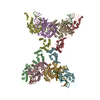
|
|---|---|
| 1 |
|
-Components
-Protein , 2 types, 8 molecules HDBFIECG
| #3: Protein | Mass: 53256.734 Da / Num. of mol.: 4 Source method: isolated from a genetically manipulated source Source: (gene. exp.) Thermoflavifilum thermophilum (bacteria)Gene: SAMN05660895_1670 / Production host: #4: Protein | Mass: 58304.848 Da / Num. of mol.: 4 Source method: isolated from a genetically manipulated source Source: (gene. exp.) Thermoflavifilum thermophilum (bacteria)Gene: SAMN05660895_1671 / Production host: |
|---|
-RNA chain / DNA chain , 2 types, 8 molecules UMSXVNTY
| #1: RNA chain | Mass: 6812.045 Da / Num. of mol.: 4 / Source method: obtained synthetically / Source: (synth.) synthetic construct (others) #2: DNA chain | Mass: 13703.841 Da / Num. of mol.: 4 / Source method: obtained synthetically / Source: (synth.) synthetic construct (others) |
|---|
-Non-polymers , 2 types, 61 molecules 

| #5: Chemical | ChemComp-MG /  Mass: 24.305 Da / Num. of mol.: 4 / Source method: obtained synthetically / Formula: Mg / Feature type: SUBJECT OF INVESTIGATION Mass: 24.305 Da / Num. of mol.: 4 / Source method: obtained synthetically / Formula: Mg / Feature type: SUBJECT OF INVESTIGATION#6: Water | ChemComp-HOH / | Mass: 18.015 Da / Num. of mol.: 57 / Source method: isolated from a natural source / Formula: H2O |
|---|
-Details
| Has ligand of interest | Y |
|---|---|
| Has protein modification | N |
-Experimental details
-Experiment
| Experiment | Method: ELECTRON MICROSCOPY |
|---|---|
| EM experiment | Aggregation state: PARTICLE / 3D reconstruction method: single particle reconstruction |
- Sample preparation
| Component | Name: SPARTA complex with gRNA/tDNA / Type: COMPLEX / Entity ID: #1-#4 / Source: MULTIPLE SOURCES |
|---|---|
| Source (natural) | Organism: Thermoflavifilum thermophilum (bacteria) |
| Source (recombinant) | Organism: |
| Buffer solution | pH: 7.5 |
| Specimen | Embedding applied: YES / Shadowing applied: NO / Staining applied: NO / Vitrification applied: YES |
| EM embedding | Material: Vitreous ice |
| Vitrification | Cryogen name: ETHANE |
- Electron microscopy imaging
| Experimental equipment |  Model: Titan Krios / Image courtesy: FEI Company |
|---|---|
| Microscopy | Model: FEI TITAN KRIOS |
| Electron gun | Electron source:  FIELD EMISSION GUN / Accelerating voltage: 300 kV / Illumination mode: FLOOD BEAM FIELD EMISSION GUN / Accelerating voltage: 300 kV / Illumination mode: FLOOD BEAM |
| Electron lens | Mode: BRIGHT FIELD / Nominal defocus max: 2500 nm / Nominal defocus min: 800 nm |
| Image recording | Electron dose: 51.11 e/Å2 / Film or detector model: GATAN K3 BIOQUANTUM (6k x 4k) |
- Processing
| EM software | Name: PHENIX / Category: model refinement | ||||||||||||||||||||||||
|---|---|---|---|---|---|---|---|---|---|---|---|---|---|---|---|---|---|---|---|---|---|---|---|---|---|
| CTF correction | Type: PHASE FLIPPING AND AMPLITUDE CORRECTION | ||||||||||||||||||||||||
| 3D reconstruction | Resolution: 3.15 Å / Resolution method: FSC 0.143 CUT-OFF / Num. of particles: 238432 / Details: Composite cryo-EM map / Symmetry type: POINT | ||||||||||||||||||||||||
| Refine LS restraints |
|
