[English] 日本語
Yorodumi
- PDB-8u7b: Crystal structure of Apo form of Short Prokaryotic Argonaute TIR-... -

+
Open data


ID or keywords:

Loading...

-
Basic information

Entry
Database: PDB / ID: 8u7b
TitleCrystal structure of Apo form of Short Prokaryotic Argonaute TIR-APAZ (SPARTA) heterodimer
Components
  • Piwi domain-containing protein
  • TIR domain-containing protein
KeywordsRNA BINDING PROTEIN / SPARTA / Short Prokaryotic Argonaute / TIR / APAZ
Function / homology
Function and homology information


nucleic acid binding / signal transduction
Similarity search - Function
TIR domain / Toll/interleukin-1 receptor homology (TIR) domain / Toll/interleukin-1 receptor homology (TIR) domain superfamily / Ribonuclease H superfamily / Ribonuclease H-like superfamily
Similarity search - Domain/homology
: / PROLINE / Piwi domain-containing protein / TIR domain-containing protein
Similarity search - Component
Biological speciesThermoflavifilum thermophilum (bacteria)
MethodX-RAY DIFFRACTION / SYNCHROTRON / MOLECULAR REPLACEMENT / Resolution: 2.66 Å
AuthorsKottur, J. / Aggarwal, A.K.
Funding support1items
OrganizationGrant numberCountry
Not funded
CitationJournal: Nat Commun / Year: 2024
Title: Nucleic acid mediated activation of a short prokaryotic Argonaute immune system.
Authors: Jithesh Kottur / Radhika Malik / Aneel K Aggarwal /
Abstract: A short prokaryotic Argonaute (pAgo) TIR-APAZ (SPARTA) defense system, activated by invading DNA to unleash its TIR domain for NAD(P) hydrolysis, was recently identified in bacteria. We report the ...A short prokaryotic Argonaute (pAgo) TIR-APAZ (SPARTA) defense system, activated by invading DNA to unleash its TIR domain for NAD(P) hydrolysis, was recently identified in bacteria. We report the crystal structure of SPARTA heterodimer in the absence of guide-RNA/target-ssDNA (2.66 Å) and a cryo-EM structure of the SPARTA oligomer (tetramer of heterodimers) bound to guide-RNA/target-ssDNA at nominal 3.15-3.35 Å resolution. The crystal structure provides a high-resolution view of SPARTA, revealing the APAZ domain as equivalent to the N, L1, and L2 regions of long pAgos and the MID domain containing a unique insertion (insert57). Cryo-EM structure reveals regions of the PIWI (loop10-9) and APAZ (helix αN) domains that reconfigure for nucleic-acid binding and decrypts regions/residues that reorganize to expose a positively charged pocket for higher-order assembly. The TIR domains amass in a parallel-strands arrangement for catalysis. We visualize SPARTA before and after RNA/ssDNA binding and uncover the basis of its active assembly leading to abortive infection.
History
DepositionSep 15, 2023Deposition site: RCSB / Processing site: RCSB
Revision 1.0Jun 12, 2024Provider: repository / Type: Initial release
Revision 1.1Jun 19, 2024Group: Database references / Category: citation / citation_author
Item: _citation.journal_volume / _citation.page_first ..._citation.journal_volume / _citation.page_first / _citation.page_last / _citation.pdbx_database_id_PubMed / _citation.title / _citation_author.identifier_ORCID

-
Structure visualization

Structure viewerMolecule:
MolmilJmol/JSmol

Downloads & links

-
Assembly

Deposited unit
B: TIR domain-containing protein
C: Piwi domain-containing protein
hetero molecules


Theoretical massNumber of molelcules
Total (without water)112,36211
Polymers111,4462
Non-polymers9169
Water1,63991
1


  • Idetical with deposited unit
  • defined by author&software
  • Evidence: gel filtration, In the gel filtration, the heterodimer eluted as a single homogenous peak, light scattering, Mass Photometry was also used to confirm the assembly
TypeNameSymmetry operationNumber
identity operation1_555x,y,z1
Buried area6550 Å2
ΔGint-51 kcal/mol
Surface area39990 Å2
MethodPISA
Unit cell
Length a, b, c (Å)197.181, 197.181, 183.407
Angle α, β, γ (deg.)90.00, 90.00, 120.00
Int Tables number179
Space group name H-MP6522

-
Components

#1: Protein TIR domain-containing protein / TIR-APAZ


Mass: 53141.648 Da / Num. of mol.: 1
Source method: isolated from a genetically manipulated source
Source: (gene. exp.) Thermoflavifilum thermophilum (bacteria)
Gene: SAMN05660895_1670 / Production host: Escherichia coli (E. coli) / References: UniProt: A0A1I7NFG5
#2: Protein Piwi domain-containing protein / Short pAgo


Mass: 58304.848 Da / Num. of mol.: 1
Source method: isolated from a genetically manipulated source
Source: (gene. exp.) Thermoflavifilum thermophilum (bacteria)
Gene: SAMN05660895_1671 / Production host: Escherichia coli BL21(DE3) (bacteria) / References: UniProt: A0A1I7NFD7
#3: Chemical
ChemComp-PRO / PROLINE


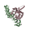
Type: L-peptide linking / Mass: 115.130 Da / Num. of mol.: 7 / Source method: obtained synthetically / Formula: C5H9NO2
#4: Chemical ChemComp-MN / MANGANESE (II) ION


Mass: 54.938 Da / Num. of mol.: 2 / Source method: obtained synthetically / Formula: Mn
#5: Water ChemComp-HOH / water


Mass: 18.015 Da / Num. of mol.: 91 / Source method: isolated from a natural source / Formula: H2O
Has ligand of interestN

-
Experimental details

-
Experiment

ExperimentMethod: X-RAY DIFFRACTION / Number of used crystals: 1

-
Sample preparation

CrystalDensity Matthews: 4.61 Å3/Da / Density % sol: 73.34 %
Crystal growTemperature: 293.15 K / Method: vapor diffusion, hanging drop / pH: 4.5
Details: 0.1 M sodium acetate, pH 4.5, 0.8 M sodium phosphate monobasic, 1.2 M potassium phosphate dibasic
PH range: 4-6

-
Data collection

DiffractionMean temperature: 100 K / Serial crystal experiment: N
Diffraction sourceSource: SYNCHROTRON / Site: NSLS-II / Beamline: 17-ID-2 / Wavelength: 0.979338 Å
DetectorType: DECTRIS EIGER X 16M / Detector: PIXEL / Date: Jul 29, 2022
RadiationProtocol: SINGLE WAVELENGTH / Monochromatic (M) / Laue (L): M / Scattering type: x-ray
Radiation wavelengthWavelength: 0.979338 Å / Relative weight: 1
ReflectionResolution: 2.66→170.76 Å / Num. obs: 40914 / % possible obs: 96.2 % / Redundancy: 13.3 % / CC1/2: 0.999 / Rmerge(I) obs: 0.148 / Rpim(I) all: 0.042 / Net I/σ(I): 13.7
Reflection shellResolution: 2.66→2.99 Å / Rmerge(I) obs: 1.767 / Mean I/σ(I) obs: 1.7 / Num. unique obs: 2047 / CC1/2: 0.572 / Rpim(I) all: 0.554

-
Processing

Software
NameVersionClassification
PHENIX(1.20rc3_4406: ???)refinement
Aimless0.7.7data scaling
XDSJan 10, 2022 (BUILT 20220110)data reduction
PHASERphasing
RefinementMethod to determine structure: MOLECULAR REPLACEMENT / Resolution: 2.66→49.71 Å / SU ML: 0.27 / Cross valid method: FREE R-VALUE / σ(F): 1.35 / Phase error: 29.72 / Stereochemistry target values: ML
RfactorNum. reflection% reflection
Rfree0.2387 2070 5.06 %
Rwork0.1793 --
obs0.1823 40895 67.56 %
Solvent computationShrinkage radii: 0.9 Å / VDW probe radii: 1.11 Å / Solvent model: FLAT BULK SOLVENT MODEL
Refinement stepCycle: LAST / Resolution: 2.66→49.71 Å
ProteinNucleic acidLigandSolventTotal
Num. atoms7365 0 51 91 7507
Refine LS restraints
Refine-IDTypeDev idealNumber
X-RAY DIFFRACTIONf_bond_d0.0037646
X-RAY DIFFRACTIONf_angle_d0.60810380
X-RAY DIFFRACTIONf_dihedral_angle_d4.9471002
X-RAY DIFFRACTIONf_chiral_restr0.0441141
X-RAY DIFFRACTIONf_plane_restr0.0041315
LS refinement shell
Resolution (Å)Rfactor RfreeNum. reflection RfreeRfactor RworkNum. reflection RworkRefine-ID% reflection obs (%)
2.66-2.720.154720.338296X-RAY DIFFRACTION2
2.72-2.790.3166130.3123228X-RAY DIFFRACTION6
2.79-2.870.322210.3218396X-RAY DIFFRACTION10
2.87-2.950.284343790X-RAY DIFFRACTION21
2.95-3.050.3497570.27231218X-RAY DIFFRACTION32
3.05-3.160.30421070.25011930X-RAY DIFFRACTION51
3.16-3.280.30261570.22393172X-RAY DIFFRACTION83
3.28-3.430.27161980.19743786X-RAY DIFFRACTION100
3.43-3.610.26392030.17523798X-RAY DIFFRACTION100
3.61-3.840.22541950.15793832X-RAY DIFFRACTION100
3.84-4.130.19641990.13753835X-RAY DIFFRACTION100
4.13-4.550.1852240.12183832X-RAY DIFFRACTION100
4.55-5.210.20942170.13373872X-RAY DIFFRACTION100
5.21-6.560.25552020.20563935X-RAY DIFFRACTION100
6.56-49.710.26472320.22714105X-RAY DIFFRACTION100

+
About Yorodumi

-
News

-
Feb 9, 2022. New format data for meta-information of EMDB entries

New format data for meta-information of EMDB entries

  • Version 3 of the EMDB header file is now the official format.
  • The previous official version 1.9 will be removed from the archive.

Related info.:EMDB header

External links:wwPDB to switch to version 3 of the EMDB data model

-
Aug 12, 2020. Covid-19 info

Covid-19 info

URL: https://pdbjlvh1.pdbj.org/emnavi/covid19.php

New page: Covid-19 featured information page in EM Navigator.

Related info.:Covid-19 info / Mar 5, 2020. Novel coronavirus structure data

+
Mar 5, 2020. Novel coronavirus structure data

Novel coronavirus structure data

Related info.:Yorodumi Speices / Aug 12, 2020. Covid-19 info

External links:COVID-19 featured content - PDBj / Molecule of the Month (242):Coronavirus Proteases

+
Jan 31, 2019. EMDB accession codes are about to change! (news from PDBe EMDB page)

EMDB accession codes are about to change! (news from PDBe EMDB page)

  • The allocation of 4 digits for EMDB accession codes will soon come to an end. Whilst these codes will remain in use, new EMDB accession codes will include an additional digit and will expand incrementally as the available range of codes is exhausted. The current 4-digit format prefixed with “EMD-” (i.e. EMD-XXXX) will advance to a 5-digit format (i.e. EMD-XXXXX), and so on. It is currently estimated that the 4-digit codes will be depleted around Spring 2019, at which point the 5-digit format will come into force.
  • The EM Navigator/Yorodumi systems omit the EMD- prefix.

Related info.:Q: What is EMD? / ID/Accession-code notation in Yorodumi/EM Navigator

External links:EMDB Accession Codes are Changing Soon! / Contact to PDBj

+
Jul 12, 2017. Major update of PDB

Major update of PDB

  • wwPDB released updated PDB data conforming to the new PDBx/mmCIF dictionary.
  • This is a major update changing the version number from 4 to 5, and with Remediation, in which all the entries are updated.
  • In this update, many items about electron microscopy experimental information are reorganized (e.g. em_software).
  • Now, EM Navigator and Yorodumi are based on the updated data.

External links:wwPDB Remediation / Enriched Model Files Conforming to OneDep Data Standards Now Available in the PDB FTP Archive

-
Yorodumi

Thousand views of thousand structures

  • Yorodumi is a browser for structure data from EMDB, PDB, SASBDB, etc.
  • This page is also the successor to EM Navigator detail page, and also detail information page/front-end page for Omokage search.
  • The word "yorodu" (or yorozu) is an old Japanese word meaning "ten thousand". "mi" (miru) is to see.

Related info.:EMDB / PDB / SASBDB / Comparison of 3 databanks / Yorodumi Search / Aug 31, 2016. New EM Navigator & Yorodumi / Yorodumi Papers / Jmol/JSmol / Function and homology information / Changes in new EM Navigator and Yorodumi

Read more