 Movie
Movie Controller

[English] 日本語
 Yorodumi
- PDB-8u26: Gaussian Mixture Models based single particle refinement - GPCR (... -
+ Open data

- Basic information
| Entry | Database: PDB / ID: 8u26 | ||||||
|---|---|---|---|---|---|---|---|
| Title | Gaussian Mixture Models based single particle refinement - GPCR (Substance P bound to active human neurokinin 1 receptor in complex with miniGs399 from EMPIAR-10786) | ||||||
 Components Components |
| ||||||
 Keywords Keywords | MEMBRANE PROTEIN / GPCR | ||||||
| Function / homology |  Function and homology information Function and homology informationsubstance P receptor binding / substance P receptor activity / positive regulation of corticosterone secretion / tachykinin receptor activity / phospholipase C-activating tachykinin receptor signaling pathway / positive regulation of flagellated sperm motility / aggressive behavior / Tachykinin receptors bind tachykinins / insemination / positive regulation of uterine smooth muscle contraction ...substance P receptor binding / substance P receptor activity / positive regulation of corticosterone secretion / tachykinin receptor activity / phospholipase C-activating tachykinin receptor signaling pathway / positive regulation of flagellated sperm motility / aggressive behavior / Tachykinin receptors bind tachykinins / insemination / positive regulation of uterine smooth muscle contraction / positive regulation of synaptic transmission, cholinergic / detection of abiotic stimulus / sperm ejaculation / tachykinin receptor signaling pathway / positive regulation of lymphocyte proliferation / sperm head / response to ozone / operant conditioning / positive regulation of action potential / positive regulation of acute inflammatory response / positive regulation of blood pressure / smooth muscle contraction involved in micturition / response to auditory stimulus / positive regulation of vascular permeability / regulation of smooth muscle cell proliferation / positive regulation of hormone secretion / regulation of smooth muscle cell migration / positive regulation of ossification / positive regulation of leukocyte migration / negative regulation of heart rate / response to pain / eating behavior / adenylate cyclase-activating G protein-coupled bile acid receptor signaling pathway / adenylate cyclase-activating serotonin receptor signaling pathway / angiotensin-mediated drinking behavior / regulation of skeletal muscle contraction / positive regulation of epithelial cell migration / behavioral response to pain / PKA activation in glucagon signalling / response to electrical stimulus / positive regulation of vasoconstriction / associative learning / hair follicle placode formation / developmental growth / intracellular transport / D1 dopamine receptor binding / sperm flagellum / neuronal dense core vesicle / long-term memory / vascular endothelial cell response to laminar fluid shear stress / renal water homeostasis / activation of adenylate cyclase activity / Hedgehog 'off' state / adenylate cyclase-activating adrenergic receptor signaling pathway / cellular response to acidic pH / positive regulation of stress fiber assembly / response to progesterone / response to hormone / cellular response to glucagon stimulus / sensory perception of pain / intracellular glucose homeostasis / positive regulation of epithelial cell proliferation / adenylate cyclase activator activity / positive regulation of insulin secretion involved in cellular response to glucose stimulus / trans-Golgi network membrane / positive regulation of synaptic transmission, GABAergic / response to nicotine / negative regulation of inflammatory response to antigenic stimulus / neuropeptide signaling pathway / response to prostaglandin E / cellular response to nerve growth factor stimulus / bone development / platelet aggregation / regulation of blood pressure / cognition / G-protein beta/gamma-subunit complex binding / positive regulation of insulin secretion / Olfactory Signaling Pathway / Activation of the phototransduction cascade / G protein-coupled acetylcholine receptor signaling pathway / G beta:gamma signalling through PLC beta / Presynaptic function of Kainate receptors / Thromboxane signalling through TP receptor / Activation of G protein gated Potassium channels / Inhibition of voltage gated Ca2+ channels via Gbeta/gamma subunits / G-protein activation / sensory perception of smell / Glucagon signaling in metabolic regulation / Prostacyclin signalling through prostacyclin receptor / G beta:gamma signalling through CDC42 / Synthesis, secretion, and inactivation of Glucagon-like Peptide-1 (GLP-1) / G beta:gamma signalling through BTK / photoreceptor disc membrane / ADP signalling through P2Y purinoceptor 12 / Sensory perception of sweet, bitter, and umami (glutamate) taste / response to estradiol / Glucagon-type ligand receptors / Adrenaline,noradrenaline inhibits insulin secretion / Vasopressin regulates renal water homeostasis via Aquaporins / Glucagon-like Peptide-1 (GLP1) regulates insulin secretion Similarity search - Function | ||||||
| Biological species |  Homo sapiens (human) Homo sapiens (human) | ||||||
| Method | ELECTRON MICROSCOPY / single particle reconstruction / cryo EM / Resolution: 2.5 Å | ||||||
 Authors Authors | Chen, M. / Pintilie, G. | ||||||
| Funding support |  United States, 1items United States, 1items
| ||||||
 Citation Citation | Journal: Nat Methods / Year: 2024 Title: Improving resolution and resolvability of single-particle cryoEM structures using Gaussian mixture models. Authors: Muyuan Chen / Michael F Schmid / Wah Chiu / Abstract: Cryogenic electron microscopy is widely used in structural biology, but its resolution is often limited by the dynamics of the macromolecule. Here we developed a refinement protocol based on Gaussian ...Cryogenic electron microscopy is widely used in structural biology, but its resolution is often limited by the dynamics of the macromolecule. Here we developed a refinement protocol based on Gaussian mixture models that integrates particle orientation and conformation estimation and improves the alignment for flexible domains of protein structures. We demonstrated this protocol on multiple datasets, resulting in improved resolution and resolvability, locally and globally, by visual and quantitative measures. | ||||||
| History |
|
- Structure visualization
| Structure viewer | Molecule: MolmilJmol/JSmol |
|---|
- Downloads & links
-Download
| PDBx/mmCIF format | 8u26.cif.gz | 461.5 KB | Display | PDBx/mmCIF format |
|---|---|---|---|---|
| PDB format | pdb8u26.ent.gz | 300.4 KB | Display | PDB format |
| PDBx/mmJSON format | 8u26.json.gz | Tree view | PDBx/mmJSON format | |
| Others |  Other downloads Other downloads |
-Validation report
| Arichive directory | https://data.pdbj.org/pub/pdb/validation_reports/u2/8u26ftp://data.pdbj.org/pub/pdb/validation_reports/u2/8u26 | HTTPS FTP |
|---|
-Related structure data
| Related structure data |  41840MC 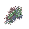 8u28C 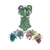 8u2cC M: map data used to model this data C: citing same article ( |
|---|---|
| Similar structure data | |
| Experimental dataset #1 | Data reference: 10.6019/EMPIAR-10786 / Data set type: EMPIAR |


-Links
 PDBj
PDBj


- Assembly
| Deposited unit | 
|
|---|---|
| 1 |
|
-Components
-Guanine nucleotide-binding protein ... , 3 types, 3 molecules ABG
| #1: Protein | Mass: 28907.684 Da / Num. of mol.: 1 Source method: isolated from a genetically manipulated source Source: (gene. exp.) Homo sapiens (human) / Gene: GNAS, GNAS1, GSP / Production host: Homo sapiens (human) / References: UniProt: P63092 |
|---|---|
| #2: Protein | Mass: 40786.566 Da / Num. of mol.: 1 Source method: isolated from a genetically manipulated source Source: (gene. exp.) Homo sapiens (human) / Gene: GNB1 / Production host: Homo sapiens (human) / References: UniProt: P62873 |
| #3: Protein | Mass: 7563.750 Da / Num. of mol.: 1 Source method: isolated from a genetically manipulated source Source: (gene. exp.) Homo sapiens (human) / Gene: GNG2 / Production host: Homo sapiens (human) / References: UniProt: P59768 |
-Antibody / Protein / Protein/peptide , 3 types, 3 molecules NRS
| #4: Antibody | Mass: 15398.067 Da / Num. of mol.: 1 Source method: isolated from a genetically manipulated source Source: (gene. exp.) |
|---|---|
| #5: Protein | Mass: 47542.867 Da / Num. of mol.: 1 Source method: isolated from a genetically manipulated source Source: (gene. exp.) Homo sapiens (human) / Gene: TACR1, NK1R, TAC1R / Production host: Homo sapiens (human) / References: UniProt: P25103 |
| #6: Protein/peptide | Mass: 1350.629 Da / Num. of mol.: 1 Source method: isolated from a genetically manipulated source Source: (gene. exp.) Homo sapiens (human) / Gene: TAC1, NKA, NKNA, TAC2 / Production host: Homo sapiens (human) / References: UniProt: P20366 |
-Details
| Has protein modification | Y |
|---|
-Experimental details
-Experiment
| Experiment | Method: ELECTRON MICROSCOPY |
|---|---|
| EM experiment | Aggregation state: PARTICLE / 3D reconstruction method: single particle reconstruction |
- Sample preparation
| Component | Name: Substance P bound to active human neurokinin 1 receptor in complex with miniGs399 Type: COMPLEX Details: Reprocessing of EMPIAR-10786. Sample information is the same as EMD-24570, PDB-7RMH. Entity ID: all / Source: NATURAL |
|---|---|
| Molecular weight | Value: 0.141 MDa / Experimental value: YES |
| Source (natural) | Organism: Homo sapiens (human) |
| Buffer solution | pH: 7.5 |
| Specimen | Embedding applied: NO / Shadowing applied: NO / Staining applied: NO / Vitrification applied: YES |
| Vitrification | Cryogen name: ETHANE |
- Electron microscopy imaging
| Experimental equipment |  Model: Titan Krios / Image courtesy: FEI Company |
|---|---|
| Microscopy | Model: FEI TITAN KRIOS |
| Electron gun | Electron source:  FIELD EMISSION GUN / Accelerating voltage: 300 kV / Illumination mode: FLOOD BEAM FIELD EMISSION GUN / Accelerating voltage: 300 kV / Illumination mode: FLOOD BEAM |
| Electron lens | Mode: BRIGHT FIELD / Nominal defocus max: 2000 nm / Nominal defocus min: 1000 nm |
| Image recording | Electron dose: 30 e/Å2 / Film or detector model: GATAN K3 (6k x 4k) |
- Processing
| EM software |
| ||||||||||||||||||||||||
|---|---|---|---|---|---|---|---|---|---|---|---|---|---|---|---|---|---|---|---|---|---|---|---|---|---|
| CTF correction | Type: PHASE FLIPPING ONLY | ||||||||||||||||||||||||
| 3D reconstruction | Resolution: 2.5 Å / Resolution method: FSC 0.143 CUT-OFF / Num. of particles: 288659 / Symmetry type: POINT | ||||||||||||||||||||||||
| Atomic model building | Protocol: FLEXIBLE FIT / Space: REAL | ||||||||||||||||||||||||
| Refine LS restraints |
|
