Movie
Movie Controller

[English] 日本語
 Yorodumi
- PDB-8tpw: Crosslinked 6-deoxyerythronolide B synthase (DEBS) Module 3 in co... -
+ Open data

- Basic information
| Entry | Database: PDB / ID: 8tpw | |||||||||||||||
|---|---|---|---|---|---|---|---|---|---|---|---|---|---|---|---|---|
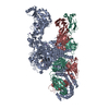
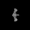
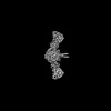
| Title | Crosslinked 6-deoxyerythronolide B synthase (DEBS) Module 3 in complex with antibody fragment 1B2: cis-oriented 1B2 and ACP | |||||||||||||||
 Components Components |
| |||||||||||||||
 Keywords Keywords | BIOSYNTHETIC PROTEIN / polyketide synthase / antibody | |||||||||||||||
| Function / homology |  Function and homology information Function and homology information6-deoxyerythronolide-B synthase / erythronolide synthase activity / macrolide biosynthetic process / fatty acid synthase activity / phosphopantetheine binding / 3-oxoacyl-[acyl-carrier-protein] synthase activity / fatty acid biosynthetic process / oxidoreductase activity Similarity search - Function | |||||||||||||||
| Biological species |  Saccharopolyspora erythraea (bacteria) Saccharopolyspora erythraea (bacteria) Homo sapiens (human) Homo sapiens (human) | |||||||||||||||
| Method | ELECTRON MICROSCOPY / single particle reconstruction / cryo EM / Resolution: 3.46 Å | |||||||||||||||
 Authors Authors | Cogan, D.P. / Soohoo, A.M. / Chen, M. / Brodsky, K.L. / Liu, Y. / Khosla, C. | |||||||||||||||
| Funding support |  United States, 4items United States, 4items
| |||||||||||||||
 Citation Citation | Journal: Nat Chem Biol / Year: 2025 Title: Structural basis for intermodular communication in assembly-line polyketide biosynthesis. Authors: Dillon P Cogan / Alexander M Soohoo / Muyuan Chen / Yan Liu / Krystal L Brodsky / Chaitan Khosla / Abstract: Assembly-line polyketide synthases (PKSs) are modular multi-enzyme systems with considerable potential for genetic reprogramming. Understanding how they selectively transport biosynthetic ...Assembly-line polyketide synthases (PKSs) are modular multi-enzyme systems with considerable potential for genetic reprogramming. Understanding how they selectively transport biosynthetic intermediates along a defined sequence of active sites could be harnessed to rationally alter PKS product structures. To investigate functional interactions between PKS catalytic and substrate acyl carrier protein (ACP) domains, we employed a bifunctional reagent to crosslink transient domain-domain interfaces of a prototypical assembly line, the 6-deoxyerythronolide B synthase, and resolved their structures by single-particle cryogenic electron microscopy (cryo-EM). Together with statistical per-particle image analysis of cryo-EM data, we uncovered interactions between ketosynthase (KS) and ACP domains that discriminate between intra-modular and inter-modular communication while reinforcing the relevance of conformational asymmetry during the catalytic cycle. Our findings provide a foundation for the structure-based design of hybrid PKSs comprising biosynthetic modules from different naturally occurring assembly lines. | |||||||||||||||
| History |
|
- Structure visualization
| Structure viewer | Molecule: MolmilJmol/JSmol |
|---|
- Downloads & links
-Download
| PDBx/mmCIF format | 8tpw.cif.gz | 492.3 KB | Display | PDBx/mmCIF format |
|---|---|---|---|---|
| PDB format | pdb8tpw.ent.gz | 356.6 KB | Display | PDB format |
| PDBx/mmJSON format | 8tpw.json.gz | Tree view | PDBx/mmJSON format | |
| Others |  Other downloads Other downloads |
-Validation report
| Arichive directory | https://data.pdbj.org/pub/pdb/validation_reports/tp/8tpwftp://data.pdbj.org/pub/pdb/validation_reports/tp/8tpw | HTTPS FTP |
|---|
-Related structure data
| Related structure data |  41495MC 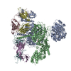 8tjnC 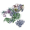 8tjoC 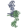 8tjpC 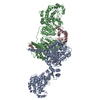 8tkoC 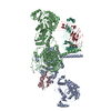 8tpxC M: map data used to model this data C: citing same article ( |
|---|---|
| Similar structure data |

-Links
 PDBj
PDBj


- Assembly
| Deposited unit | 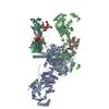
|
|---|---|
| 1 |
|
-Components
| #1: Protein | Mass: 186489.578 Da / Num. of mol.: 3 Fragment: DEBS Module 3,DEBS Module 3,DEBS Module 3,DEBS Module 3 Source method: isolated from a genetically manipulated source Source: (gene. exp.) Saccharopolyspora erythraea (bacteria) / Gene: eryAII, eryA / Plasmid: pRSG34 / Production host: References: UniProt: Q5UNP5, 6-deoxyerythronolide-B synthase #2: Antibody | | Mass: 26447.611 Da / Num. of mol.: 1 Source method: isolated from a genetically manipulated source Source: (gene. exp.) Homo sapiens (human) / Production host: #3: Antibody | | Mass: 25715.832 Da / Num. of mol.: 1 Source method: isolated from a genetically manipulated source Source: (gene. exp.) Homo sapiens (human) / Production host: Has ligand of interest | Y | Has protein modification | Y | |
|---|
-Experimental details
-Experiment
| Experiment | Method: ELECTRON MICROSCOPY |
|---|---|
| EM experiment | Aggregation state: PARTICLE / 3D reconstruction method: single particle reconstruction |
- Sample preparation
| Component | Name: Crosslinked DEBS Module 3 in complex with Antibody Fragment 1B2 Type: COMPLEX / Entity ID: all / Source: MULTIPLE SOURCES | ||||||||||||||||||||
|---|---|---|---|---|---|---|---|---|---|---|---|---|---|---|---|---|---|---|---|---|---|
| Molecular weight | Value: 0.42 MDa / Experimental value: YES | ||||||||||||||||||||
| Source (natural) | Organism: Saccharopolyspora erythraea (bacteria) | ||||||||||||||||||||
| Source (recombinant) | Organism: | ||||||||||||||||||||
| Buffer solution | pH: 7.2 | ||||||||||||||||||||
| Buffer component |
| ||||||||||||||||||||
| Specimen | Conc.: 8 mg/ml / Embedding applied: NO / Shadowing applied: NO / Staining applied: NO / Vitrification applied: YES | ||||||||||||||||||||
| Specimen support | Details: 30 s glow, 10 s hold, 15 mA / Grid material: COPPER / Grid mesh size: 300 divisions/in. / Grid type: Quantifoil R2/1 | ||||||||||||||||||||
| Vitrification | Instrument: FEI VITROBOT MARK IV / Cryogen name: ETHANE / Humidity: 100 % / Chamber temperature: 277.15 K |
- Electron microscopy imaging
| Experimental equipment |  Model: Titan Krios / Image courtesy: FEI Company |
|---|---|
| Microscopy | Model: FEI TITAN KRIOS |
| Electron gun | Electron source:  FIELD EMISSION GUN / Accelerating voltage: 300 kV / Illumination mode: FLOOD BEAM FIELD EMISSION GUN / Accelerating voltage: 300 kV / Illumination mode: FLOOD BEAM |
| Electron lens | Mode: BRIGHT FIELD / Nominal magnification: 81000 X / Nominal defocus max: 2500 nm / Nominal defocus min: 1000 nm / Cs: 2.7 mm / C2 aperture diameter: 150 µm |
| Specimen holder | Cryogen: NITROGEN / Specimen holder model: FEI TITAN KRIOS AUTOGRID HOLDER |
| Image recording | Average exposure time: 2.79 sec. / Electron dose: 50 e/Å2 / Film or detector model: GATAN K3 BIOQUANTUM (6k x 4k) / Num. of grids imaged: 1 / Num. of real images: 10480 |
- Processing
| EM software | Name: PHENIX / Version: 1.21_5207 / Category: model refinement |
|---|---|
| CTF correction | Type: PHASE FLIPPING AND AMPLITUDE CORRECTION |
| Particle selection | Num. of particles selected: 573745 |
| Symmetry | Point symmetry: C1 (asymmetric) |
| 3D reconstruction | Resolution: 3.46 Å / Resolution method: FSC 0.143 CUT-OFF / Num. of particles: 59205 / Num. of class averages: 1 / Symmetry type: POINT |
| Atomic model building | Protocol: RIGID BODY FIT / Space: REAL |
| Atomic model building | PDB-ID: 6C9U Accession code: 6C9U / Source name: PDB / Type: experimental model |
| Refinement | Cross valid method: NONE |