 Movie
Movie Controller


+ Open data

- Basic information
| Entry | Database: PDB / ID: 8r51 | ||||||
|---|---|---|---|---|---|---|---|
| Title | Mouse teneurin-3 non-compact subunit - A1B1 isoform | ||||||
 Components Components | Teneurin-3 | ||||||
 Keywords Keywords | CELL ADHESION / Synaptic cell adhesion molecule / Homodimer / Cis-synaptic | ||||||
| Function / homology |  Function and homology information Function and homology informationregulation of homophilic cell adhesion / synaptic membrane adhesion / heterophilic cell-cell adhesion via plasma membrane cell adhesion molecules / homophilic cell adhesion via plasma membrane adhesion molecules / neuron development / cell adhesion molecule binding / presynaptic active zone membrane / positive regulation of neuron projection development / neuron projection / protein heterodimerization activity ...regulation of homophilic cell adhesion / synaptic membrane adhesion / heterophilic cell-cell adhesion via plasma membrane cell adhesion molecules / homophilic cell adhesion via plasma membrane adhesion molecules / neuron development / cell adhesion molecule binding / presynaptic active zone membrane / positive regulation of neuron projection development / neuron projection / protein heterodimerization activity / axon / glutamatergic synapse / signal transduction / protein homodimerization activity / identical protein binding / membrane / plasma membrane Similarity search - Function | ||||||
| Biological species |  | ||||||
| Method | ELECTRON MICROSCOPY / single particle reconstruction / cryo EM / Resolution: 3.2 Å | ||||||
 Authors Authors | Gogou, C. / Meijer, D.H. | ||||||
| Funding support |  Netherlands, 1items Netherlands, 1items
| ||||||
 Citation Citation | Journal: Nat Commun / Year: 2024 Title: Alternative splicing controls teneurin-3 compact dimer formation for neuronal recognition. Authors: Christos Gogou / J Wouter Beugelink / Cátia P Frias / Leanid Kresik / Natalia Jaroszynska / Uwe Drescher / Bert J C Janssen / Robert Hindges / Dimphna H Meijer /  Abstract: Neuronal network formation is facilitated by recognition between synaptic cell adhesion molecules at the cell surface. Alternative splicing of cell adhesion molecules provides additional specificity ...Neuronal network formation is facilitated by recognition between synaptic cell adhesion molecules at the cell surface. Alternative splicing of cell adhesion molecules provides additional specificity in forming neuronal connections. For the teneurin family of cell adhesion molecules, alternative splicing of the EGF-repeats and NHL domain controls synaptic protein-protein interactions. Here we present cryo-EM structures of the compact dimeric ectodomain of two teneurin-3 isoforms that harbour the splice insert in the EGF-repeats. This dimer is stabilised by an EGF8-ABD contact between subunits. Cryo-EM reconstructions of all four splice variants, together with SAXS and negative stain EM, reveal compacted dimers for each, with variant-specific dimeric arrangements. This results in specific trans-cellular interactions, as tested in cell clustering and stripe assays. The compact conformations provide a structural basis for teneurin homo- and heterophilic interactions. Altogether, our findings demonstrate how alternative splicing results in rearrangements of the dimeric subunits, influencing neuronal recognition and likely circuit wiring. #1: Journal: BioRxivTitle: Mouse teneurin-3 non-compact subunit - A1B1 isoform Authors: Gogou, C. / Meijer, D.H. | ||||||
| History |
|
- Structure visualization
| Structure viewer | Molecule: MolmilJmol/JSmol |
|---|
- Downloads & links
-Download
| PDBx/mmCIF format | 8r51.cif.gz | 345.4 KB | Display | PDBx/mmCIF format |
|---|---|---|---|---|
| PDB format | pdb8r51.ent.gz | 259.2 KB | Display | PDB format |
| PDBx/mmJSON format | 8r51.json.gz | Tree view | PDBx/mmJSON format | |
| Others |  Other downloads Other downloads |
-Validation report
| Summary document | 8r51_validation.pdf.gz | 1.5 MB | Display | wwPDB validaton report |
|---|---|---|---|---|
| Full document | 8r51_full_validation.pdf.gz | 1.5 MB | Display | |
| Data in XML | 8r51_validation.xml.gz | 59.4 KB | Display | |
| Data in CIF | 8r51_validation.cif.gz | 89.2 KB | Display | |
| Arichive directory | https://data.pdbj.org/pub/pdb/validation_reports/r5/8r51ftp://data.pdbj.org/pub/pdb/validation_reports/r5/8r51 | HTTPS FTP |
-Related structure data
| Related structure data | 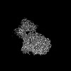 18890MC 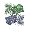 8r50C  8r54C M: map data used to model this data C: citing same article ( |
|---|---|
| Similar structure data | |
| Other databases |
|


-Links
 PDBj
PDBj


- Assembly
| Deposited unit | 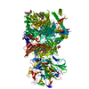
|
|---|---|
| 1 |
|
-Components
| #1: Protein | Mass: 268722.250 Da / Num. of mol.: 1 Source method: isolated from a genetically manipulated source Source: (gene. exp.)  Homo sapiens (human) / References: UniProt: Q9WTS6 Homo sapiens (human) / References: UniProt: Q9WTS6 | ||||
|---|---|---|---|---|---|
| #2: Polysaccharide | 2-acetamido-2-deoxy-beta-D-glucopyranose-(1-4)-2-acetamido-2-deoxy-beta-D-glucopyranose Source method: isolated from a genetically manipulated source #3: Sugar | ChemComp-NAG /   Type: D-saccharide, beta linking / Mass: 221.208 Da / Num. of mol.: 5 / Source method: obtained synthetically / Formula: C8H15NO6 Type: D-saccharide, beta linking / Mass: 221.208 Da / Num. of mol.: 5 / Source method: obtained synthetically / Formula: C8H15NO6Has ligand of interest | N | |
-Experimental details
-Experiment
| Experiment | Method: ELECTRON MICROSCOPY |
|---|---|
| EM experiment | Aggregation state: PARTICLE / 3D reconstruction method: single particle reconstruction |
- Sample preparation
| Component | Name: mouse teneurin-3 A1B1 isoform ectodomain subunit / Type: COMPLEX / Entity ID: #1 / Source: RECOMBINANT |
|---|---|
| Molecular weight | Value: 0.53696 MDa / Experimental value: NO |
| Source (natural) | Organism: |
| Source (recombinant) | Organism: Homo sapiens (human) / Cell: embryonic kidney |
| Buffer solution | pH: 7.8 / Details: 20 mM HEPES, 150 mM NaCl, 2mM CaCl2 |
| Specimen | Conc.: 0.5 mg/ml / Embedding applied: NO / Shadowing applied: NO / Staining applied: NO / Vitrification applied: YES |
| Vitrification | Cryogen name: ETHANE / Humidity: 100 % / Chamber temperature: 22 K |
- Electron microscopy imaging
| Experimental equipment |  Model: Titan Krios / Image courtesy: FEI Company |
|---|---|
| Microscopy | Model: TFS KRIOS |
| Electron gun | Electron source:  FIELD EMISSION GUN / Accelerating voltage: 300 kV / Illumination mode: SPOT SCAN FIELD EMISSION GUN / Accelerating voltage: 300 kV / Illumination mode: SPOT SCAN |
| Electron lens | Mode: BRIGHT FIELD / Nominal defocus max: 2000 nm / Nominal defocus min: 800 nm |
| Image recording | Electron dose: 50 e/Å2 / Film or detector model: GATAN K3 (6k x 4k) |
- Processing
| CTF correction | Type: NONE | ||||||||||||||||||||||||
|---|---|---|---|---|---|---|---|---|---|---|---|---|---|---|---|---|---|---|---|---|---|---|---|---|---|
| 3D reconstruction | Resolution: 3.2 Å / Resolution method: FSC 0.143 CUT-OFF / Num. of particles: 69290 / Symmetry type: POINT | ||||||||||||||||||||||||
| Refine LS restraints |
|
