+ データを開く
- 基本情報
基本情報
| 登録情報 | データベース: PDB / ID: 8p4r | ||||||
|---|---|---|---|---|---|---|---|
| タイトル | In situ structure average of GroEL14-GroES14 complexes in Escherichia coli cytosol obtained by cryo electron tomography | ||||||
 要素 要素 |
| ||||||
 キーワード キーワード | CHAPERONE / Chaperonin / Folding cage / proteostasis / heat shock / ATPase | ||||||
| 機能・相同性 |  機能・相同性情報 機能・相同性情報GroEL-GroES complex / chaperonin ATPase / chaperone cofactor-dependent protein refolding / protein folding chaperone / isomerase activity / ATP-dependent protein folding chaperone / unfolded protein binding / protein-folding chaperone binding / response to heat / protein refolding ...GroEL-GroES complex / chaperonin ATPase / chaperone cofactor-dependent protein refolding / protein folding chaperone / isomerase activity / ATP-dependent protein folding chaperone / unfolded protein binding / protein-folding chaperone binding / response to heat / protein refolding / ATP binding / metal ion binding / cytoplasm 類似検索 - 分子機能 | ||||||
| 生物種 |  | ||||||
| 手法 | 電子顕微鏡法 / サブトモグラム平均法 / クライオ電子顕微鏡法 / 解像度: 11.9 Å | ||||||
 データ登録者 データ登録者 | Wagner, J. / Caravajal, A.I. / Beck, F. / Bracher, A. / Wan, W. / Bohn, S. / Koerner, R. / Baumeister, W. / Fernandez-Busnadiego, R. / Hartl, F.U. | ||||||
| 資金援助 | 1件
| ||||||
 引用 引用 |  ジャーナル: Nature / 年: 2024 ジャーナル: Nature / 年: 2024タイトル: Visualizing chaperonin function in situ by cryo-electron tomography. 著者: Jonathan Wagner / Alonso I Carvajal / Andreas Bracher / Florian Beck / William Wan / Stefan Bohn / Roman Körner / Wolfgang Baumeister / Ruben Fernandez-Busnadiego / F Ulrich Hartl /   要旨: Chaperonins are large barrel-shaped complexes that mediate ATP-dependent protein folding. The bacterial chaperonin GroEL forms juxtaposed rings that bind unfolded protein and the lid-shaped cofactor ...Chaperonins are large barrel-shaped complexes that mediate ATP-dependent protein folding. The bacterial chaperonin GroEL forms juxtaposed rings that bind unfolded protein and the lid-shaped cofactor GroES at their apertures. In vitro analyses of the chaperonin reaction have shown that substrate protein folds, unimpaired by aggregation, while transiently encapsulated in the GroEL central cavity by GroES. To determine the functional stoichiometry of GroEL, GroES and client protein in situ, here we visualized chaperonin complexes in their natural cellular environment using cryo-electron tomography. We find that, under various growth conditions, around 55-70% of GroEL binds GroES asymmetrically on one ring, with the remainder populating symmetrical complexes. Bound substrate protein is detected on the free ring of the asymmetrical complex, defining the substrate acceptor state. In situ analysis of GroEL-GroES chambers, validated by high-resolution structures obtained in vitro, showed the presence of encapsulated substrate protein in a folded state before release into the cytosol. Based on a comprehensive quantification and conformational analysis of chaperonin complexes, we propose a GroEL-GroES reaction cycle that consists of linked asymmetrical and symmetrical subreactions mediating protein folding. Our findings illuminate the native conformational and functional chaperonin cycle directly within cells. | ||||||
| 履歴 |
|
- 構造の表示
構造の表示
| 構造ビューア | 分子:  Molmil Molmil Jmol/JSmol Jmol/JSmol |
|---|
- ダウンロードとリンク
- ダウンロード
| PDBx/mmCIF形式 |  8p4r.cif.gz 8p4r.cif.gz | 1.3 MB | 表示 |  PDBx/mmCIF形式 PDBx/mmCIF形式 |
|---|---|---|---|---|
| PDB形式 |  pdb8p4r.ent.gz pdb8p4r.ent.gz | 1.1 MB | 表示 |  PDB形式 PDB形式 |
| PDBx/mmJSON形式 |  8p4r.json.gz 8p4r.json.gz | ツリー表示 |  PDBx/mmJSON形式 PDBx/mmJSON形式 | |
| その他 |  その他のダウンロード その他のダウンロード |
-検証レポート
| 文書・要旨 |  8p4r_validation.pdf.gz 8p4r_validation.pdf.gz | 2.2 MB | 表示 |  wwPDB検証レポート wwPDB検証レポート |
|---|---|---|---|---|
| 文書・詳細版 |  8p4r_full_validation.pdf.gz 8p4r_full_validation.pdf.gz | 2.4 MB | 表示 | |
| XML形式データ |  8p4r_validation.xml.gz 8p4r_validation.xml.gz | 215.4 KB | 表示 | |
| CIF形式データ |  8p4r_validation.cif.gz 8p4r_validation.cif.gz | 328.1 KB | 表示 | |
| アーカイブディレクトリ |  https://data.pdbj.org/pub/pdb/validation_reports/p4/8p4r https://data.pdbj.org/pub/pdb/validation_reports/p4/8p4r ftp://data.pdbj.org/pub/pdb/validation_reports/p4/8p4r ftp://data.pdbj.org/pub/pdb/validation_reports/p4/8p4r | HTTPS FTP |
-関連構造データ
| 関連構造データ |  17426MC 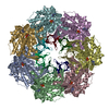 8p4mC 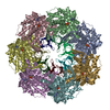 8p4nC 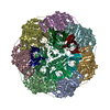 8p4oC 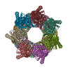 8p4pC 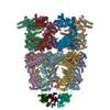 8qxsC 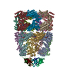 8qxtC 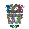 8qxuC 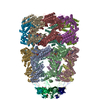 8qxvC M: このデータのモデリングに利用したマップデータ C: 同じ文献を引用 ( |
|---|---|
| 類似構造データ | 類似検索 - 機能・相同性  F&H 検索 F&H 検索 |
- リンク
リンク
- 集合体
集合体
| 登録構造単位 | 
|
|---|---|
| 1 |
|
- 要素
要素
-タンパク質 , 2種, 28分子 ABCDEFGHIJKLMNOPQRSTUVWXYZab
| #1: タンパク質 | 分子量: 57260.504 Da / 分子数: 14 / 由来タイプ: 組換発現 由来: (組換発現)  遺伝子: groEL / 発現宿主:  #2: タンパク質 | 分子量: 10400.938 Da / 分子数: 14 / 由来タイプ: 組換発現 由来: (組換発現)  遺伝子: groES / 発現宿主:  |
|---|
-非ポリマー , 4種, 448分子 


| #3: 化合物 | ChemComp-ATP / #4: 化合物 | ChemComp-MG / #5: 化合物 | ChemComp-K / #6: 水 | ChemComp-HOH / | |
|---|
-詳細
| 研究の焦点であるリガンドがあるか | N |
|---|
-実験情報
-実験
| 実験 | 手法: 電子顕微鏡法 |
|---|---|
| EM実験 | 試料の集合状態: CELL / 3次元再構成法: サブトモグラム平均法 |
- 試料調製
試料調製
| 構成要素 | 名称: GroEL14-GroES14 complex / タイプ: COMPLEX / Entity ID: #1-#2 / 由来: RECOMBINANT |
|---|---|
| 分子量 | 実験値: NO |
| 由来(天然) | 生物種:  |
| 由来(組換発現) | 生物種:  |
| 緩衝液 | pH: 7.4 |
| 試料 | 包埋: NO / シャドウイング: NO / 染色: NO / 凍結: YES / 詳細: Vitrified E. coli Bl21 (DE3) cells |
| 急速凍結 | 凍結剤: ETHANE-PROPANE |
- 電子顕微鏡撮影
| 実験機器 |  モデル: Titan Krios / 画像提供: FEI Company |
|---|---|
| 顕微鏡 | モデル: FEI TITAN KRIOS |
| 電子銃 | 電子線源:  FIELD EMISSION GUN / 加速電圧: 300 kV / 照射モード: FLOOD BEAM FIELD EMISSION GUN / 加速電圧: 300 kV / 照射モード: FLOOD BEAM |
| 電子レンズ | モード: BRIGHT FIELD / 最大 デフォーカス(公称値): 5000 nm / 最小 デフォーカス(公称値): 2500 nm / Cs: 2.7 mm / C2レンズ絞り径: 70 µm |
| 撮影 | 電子線照射量: 3 e/Å2 / Avg electron dose per subtomogram: 120 e/Å2 / 検出モード: SUPER-RESOLUTION フィルム・検出器のモデル: GATAN K2 SUMMIT (4k x 4k) |
- 解析
解析
| EMソフトウェア |
| ||||||||||||||||||||||||
|---|---|---|---|---|---|---|---|---|---|---|---|---|---|---|---|---|---|---|---|---|---|---|---|---|---|
| CTF補正 | タイプ: PHASE FLIPPING AND AMPLITUDE CORRECTION | ||||||||||||||||||||||||
| 対称性 | 点対称性: D7 (2回x7回 2面回転対称) | ||||||||||||||||||||||||
| 3次元再構成 | 解像度: 11.9 Å / 解像度の算出法: FSC 0.143 CUT-OFF / 粒子像の数: 11213 / 対称性のタイプ: POINT | ||||||||||||||||||||||||
| EM volume selection | Num. of tomograms: 216 / Num. of volumes extracted: 125860 | ||||||||||||||||||||||||
| 精密化 | 交差検証法: NONE 立体化学のターゲット値: GeoStd + Monomer Library + CDL v1.2 | ||||||||||||||||||||||||
| 原子変位パラメータ | Biso mean: 992.53 Å2 | ||||||||||||||||||||||||
| 拘束条件 |
|
 ムービー
ムービー コントローラー


 PDBj
PDBj


