 Movie
Movie Controller


+ Open data

- Basic information
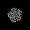
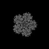
| Entry | Database: PDB / ID: 8g6m | ||||||||||||||||||
|---|---|---|---|---|---|---|---|---|---|---|---|---|---|---|---|---|---|---|---|
| Title | HIV-1 CA lattice bound to IP6, pH 7.4 | ||||||||||||||||||
 Components Components | Capsid protein | ||||||||||||||||||
 Keywords Keywords | VIRUS LIKE PARTICLE / HIV-1 / capsid / lattice / virus | ||||||||||||||||||
| Function / homology |  Function and homology information Function and homology informationviral nucleocapsid / host cell cytoplasm / viral translational frameshifting / host cell nucleus / structural molecule activity / virion membrane / RNA binding / zinc ion binding / ATP binding / cytoplasm Similarity search - Function | ||||||||||||||||||
| Biological species |   Human immunodeficiency virus 1 Human immunodeficiency virus 1 | ||||||||||||||||||
| Method | ELECTRON MICROSCOPY / single particle reconstruction / cryo EM / Resolution: 3.1 Å | ||||||||||||||||||
 Authors Authors | Highland, C.M. / Dick, R.A. | ||||||||||||||||||
| Funding support |  United States, United States,  United Kingdom, 5items United Kingdom, 5items
| ||||||||||||||||||
 Citation Citation | Journal: Proc Natl Acad Sci U S A / Year: 2023 Title: Structural insights into HIV-1 polyanion-dependent capsid lattice formation revealed by single particle cryo-EM. Authors: Carolyn M Highland / Aaron Tan / Clifton L Ricaña / John A G Briggs / Robert A Dick /  Abstract: The HIV-1 capsid houses the viral genome and interacts extensively with host cell proteins throughout the viral life cycle. It is composed of capsid protein (CA), which assembles into a conical ...The HIV-1 capsid houses the viral genome and interacts extensively with host cell proteins throughout the viral life cycle. It is composed of capsid protein (CA), which assembles into a conical fullerene lattice composed of roughly 200 CA hexamers and 12 CA pentamers. Previous structural analyses of individual CA hexamers and pentamers have provided valuable insight into capsid structure and function, but detailed structural information about these assemblies in the broader context of the capsid lattice is lacking. In this study, we combined cryoelectron tomography and single particle analysis (SPA) cryoelectron microscopy to determine structures of continuous regions of the capsid lattice containing both hexamers and pentamers. We also developed a method of liposome scaffold-based in vitro lattice assembly ("lattice templating") that enabled us to directly study the lattice under a wider range of conditions than has previously been possible. Using this approach, we identified a critical role for inositol hexakisphosphate in pentamer formation and determined the structure of the CA lattice bound to the capsid-targeting antiretroviral drug GS-6207 (lenacapavir). Our work reveals key structural details of the mature HIV-1 CA lattice and establishes the combination of lattice templating and SPA as a robust strategy for studying retroviral capsid structure and capsid interactions with host proteins and antiviral compounds. | ||||||||||||||||||
| History |
|
- Structure visualization
| Structure viewer | Molecule: MolmilJmol/JSmol |
|---|
- Downloads & links
-Download
| PDBx/mmCIF format | 8g6m.cif.gz | 493.4 KB | Display | PDBx/mmCIF format |
|---|---|---|---|---|
| PDB format | pdb8g6m.ent.gz | 415.9 KB | Display | PDB format |
| PDBx/mmJSON format | 8g6m.json.gz | Tree view | PDBx/mmJSON format | |
| Others |  Other downloads Other downloads |
-Validation report
| Summary document | 8g6m_validation.pdf.gz | 1.1 MB | Display | wwPDB validaton report |
|---|---|---|---|---|
| Full document | 8g6m_full_validation.pdf.gz | 1.1 MB | Display | |
| Data in XML | 8g6m_validation.xml.gz | 51.3 KB | Display | |
| Data in CIF | 8g6m_validation.cif.gz | 77.8 KB | Display | |
| Arichive directory | https://data.pdbj.org/pub/pdb/validation_reports/g6/8g6mftp://data.pdbj.org/pub/pdb/validation_reports/g6/8g6m | HTTPS FTP |
-Related structure data
| Related structure data |  29774MC 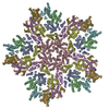 8g6kC 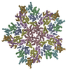 8g6lC 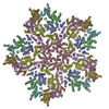 8g6nC 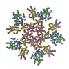 8g6oC M: map data used to model this data C: citing same article ( |
|---|---|
| Similar structure data |


-Links
 PDBj
PDBj
- Assembly
| Deposited unit | 
|
|---|---|
| 1 | x 5
|
| 2 |
|
| 3 | 
|
-Components
| #1: Protein | Mass: 26590.506 Da / Num. of mol.: 7 Source method: isolated from a genetically manipulated source Source: (gene. exp.) Human immunodeficiency virus 1 / Gene: gag / Production host:  |
|---|
-Experimental details
-Experiment
| Experiment | Method: ELECTRON MICROSCOPY |
|---|---|
| EM experiment | Aggregation state: PARTICLE / 3D reconstruction method: single particle reconstruction |
- Sample preparation
| Component | Name: HIV-1 CA lattice bound to IP6 at pH 7.4 / Type: COMPLEX Details: Highly-curved HIV-1 capsid (CA) lattice assembled on small unilamellar vesicles with inositol hexakisphosphate (IP6) at pH 7.4 Entity ID: all / Source: RECOMBINANT |
|---|---|
| Molecular weight | Experimental value: NO |
| Source (natural) | Organism: Human immunodeficiency virus 1 |
| Source (recombinant) | Organism: |
| Buffer solution | pH: 7.4 |
| Specimen | Embedding applied: NO / Shadowing applied: NO / Staining applied: NO / Vitrification applied: YES |
| Vitrification | Cryogen name: ETHANE |
- Electron microscopy imaging
| Experimental equipment |  Model: Talos Arctica / Image courtesy: FEI Company |
|---|---|
| Microscopy | Model: FEI TALOS ARCTICA |
| Electron gun | Electron source:  FIELD EMISSION GUN / Accelerating voltage: 200 kV / Illumination mode: FLOOD BEAM FIELD EMISSION GUN / Accelerating voltage: 200 kV / Illumination mode: FLOOD BEAM |
| Electron lens | Mode: BRIGHT FIELD / Nominal defocus max: 1600 nm / Nominal defocus min: 600 nm |
| Image recording | Electron dose: 50 e/Å2 / Film or detector model: GATAN K3 BIOQUANTUM (6k x 4k) |
- Processing
| Software | Name: PHENIX / Version: 1.20.1_4487: / Classification: refinement | ||||||||||||||||||||||||
|---|---|---|---|---|---|---|---|---|---|---|---|---|---|---|---|---|---|---|---|---|---|---|---|---|---|
| CTF correction | Type: PHASE FLIPPING AND AMPLITUDE CORRECTION | ||||||||||||||||||||||||
| 3D reconstruction | Resolution: 3.1 Å / Resolution method: FSC 0.143 CUT-OFF / Num. of particles: 84603 / Symmetry type: POINT | ||||||||||||||||||||||||
| Refine LS restraints |
|
