National Institutes of Health/National Institute Of Allergy and Infectious Diseases (NIH/NIAID)
R01AI147890
United States
National Institutes of Health/National Institute Of Allergy and Infectious Diseases (NIH/NIAID)
5U54AI150472-09
United States
National Institutes of Health/National Institute Of Allergy and Infectious Diseases (NIH/NIAID)
U54AI170855-01
United States
National Science Foundation (NSF, United States)
DMR-1719875
United States
Medical Research Council (MRC, United Kingdom)
MC_UP_1201/16
United Kingdom
Citation
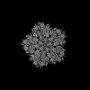
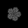
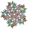
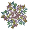
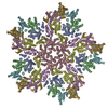
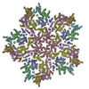
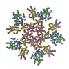
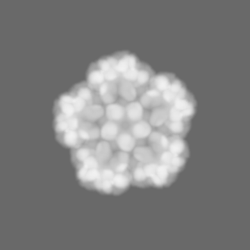
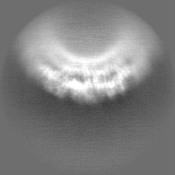
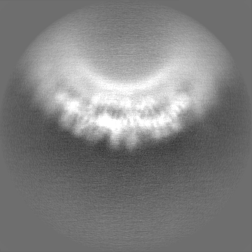
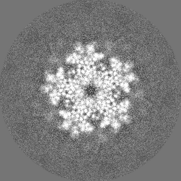
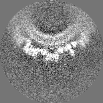
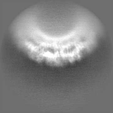
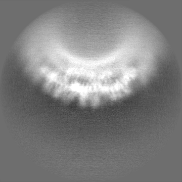
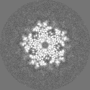
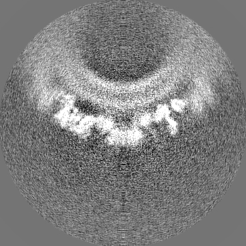
Journal: Proc Natl Acad Sci U S A / Year: 2023 Title: Structural insights into HIV-1 polyanion-dependent capsid lattice formation revealed by single particle cryo-EM. Authors: Carolyn M Highland / Aaron Tan / Clifton L Ricaña / John A G Briggs / Robert A Dick / Abstract: The HIV-1 capsid houses the viral genome and interacts extensively with host cell proteins throughout the viral life cycle. It is composed of capsid protein (CA), which assembles into a conical ...The HIV-1 capsid houses the viral genome and interacts extensively with host cell proteins throughout the viral life cycle. It is composed of capsid protein (CA), which assembles into a conical fullerene lattice composed of roughly 200 CA hexamers and 12 CA pentamers. Previous structural analyses of individual CA hexamers and pentamers have provided valuable insight into capsid structure and function, but detailed structural information about these assemblies in the broader context of the capsid lattice is lacking. In this study, we combined cryoelectron tomography and single particle analysis (SPA) cryoelectron microscopy to determine structures of continuous regions of the capsid lattice containing both hexamers and pentamers. We also developed a method of liposome scaffold-based in vitro lattice assembly ("lattice templating") that enabled us to directly study the lattice under a wider range of conditions than has previously been possible. Using this approach, we identified a critical role for inositol hexakisphosphate in pentamer formation and determined the structure of the CA lattice bound to the capsid-targeting antiretroviral drug GS-6207 (lenacapavir). Our work reveals key structural details of the mature HIV-1 CA lattice and establishes the combination of lattice templating and SPA as a robust strategy for studying retroviral capsid structure and capsid interactions with host proteins and antiviral compounds.
In the structure databanks used in Yorodumi, some data are registered as the other names, "COVID-19 virus" and "2019-nCoV". Here are the details of the virus and the list of structure data.
Jan 31, 2019. EMDB accession codes are about to change! (news from PDBe EMDB page)
EMDB accession codes are about to change! (news from PDBe EMDB page)
The allocation of 4 digits for EMDB accession codes will soon come to an end. Whilst these codes will remain in use, new EMDB accession codes will include an additional digit and will expand incrementally as the available range of codes is exhausted. The current 4-digit format prefixed with “EMD-” (i.e. EMD-XXXX) will advance to a 5-digit format (i.e. EMD-XXXXX), and so on. It is currently estimated that the 4-digit codes will be depleted around Spring 2019, at which point the 5-digit format will come into force.
The EM Navigator/Yorodumi systems omit the EMD- prefix.
Related info.:Q: What is EMD? / ID/Accession-code notation in Yorodumi/EM Navigator
Yorodumi is a browser for structure data from EMDB, PDB, SASBDB, etc.
This page is also the successor to EM Navigator detail page, and also detail information page/front-end page for Omokage search.
The word "yorodu" (or yorozu) is an old Japanese word meaning "ten thousand". "mi" (miru) is to see.
Related info.:EMDB / PDB / SASBDB / Comparison of 3 databanks / Yorodumi Search / Aug 31, 2016. New EM Navigator & Yorodumi / Yorodumi Papers / Jmol/JSmol / Function and homology information / Changes in new EM Navigator and Yorodumi
 Movie
Movie Controller


 Open data
 Basic information
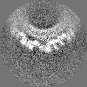
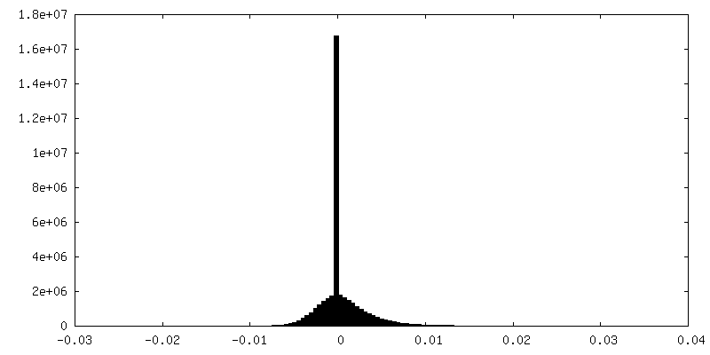
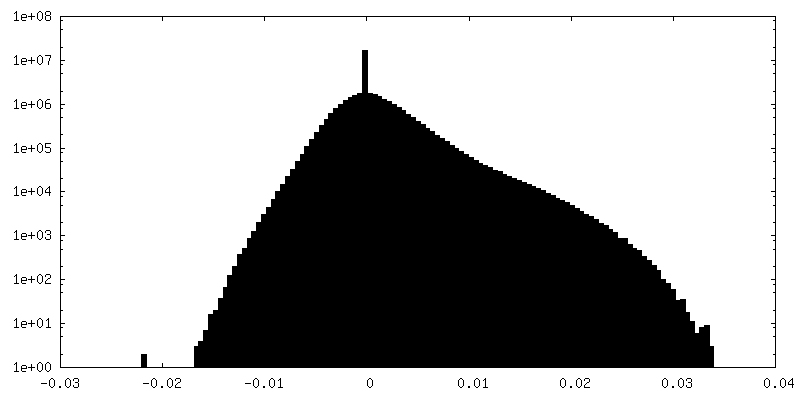
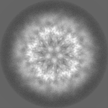
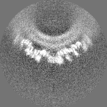
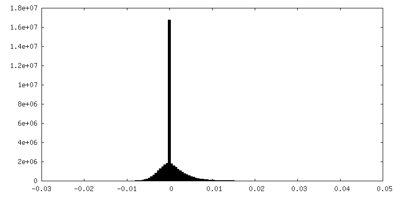
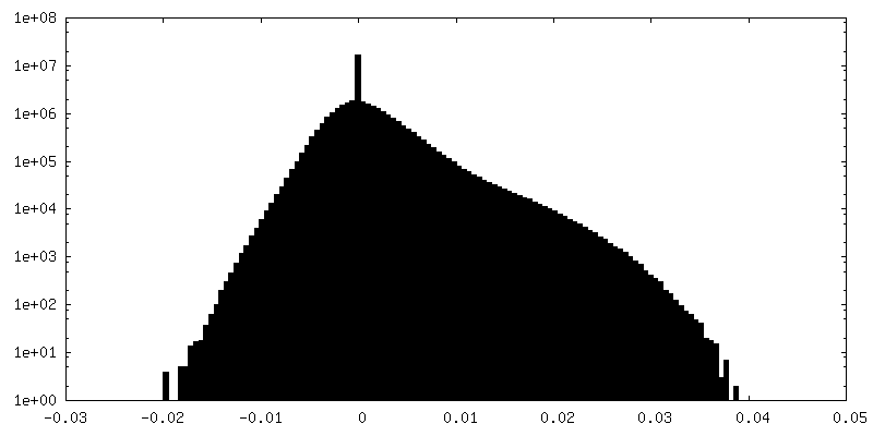
 Map data
 Sample
 Human immunodeficiency virus 1
 Authors
 United States,
  United Kingdom, 5 items
  Citation
 Structure visualization
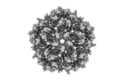
 Downloads & links
 EMDB map data format
 emd_29777.png
 http://ftp.pdbj.org/pub/emdb/structures/EMD-29777


 Z (Sec.)
 Y (Row.)
 X (Col.)


 Sample components
 Processing
 Electron microscopy
 FIELD EMISSION GUN
