 Movie
Movie Controller

[English] 日本語
 Yorodumi
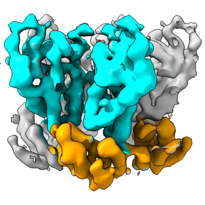
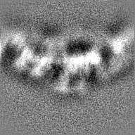
- EMDB-16698: Subtomogram average of HIV-1 CA pentamer from capsid-like particl... -
+ Open data

- Basic information
| Entry |  | |||||||||||||||||||||||||||
|---|---|---|---|---|---|---|---|---|---|---|---|---|---|---|---|---|---|---|---|---|---|---|---|---|---|---|---|---|
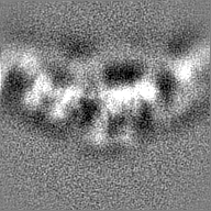
| Title | Subtomogram average of HIV-1 CA pentamer from capsid-like particles assembled with inositol hexakisphosphate | |||||||||||||||||||||||||||
 Map data Map data | Subtomogram average of HIV-1 CA protein pentamer from capsid-like particles assembled with inositol hexakisphosphate | |||||||||||||||||||||||||||
 Sample Sample |
| |||||||||||||||||||||||||||
| Biological species |  | |||||||||||||||||||||||||||
| Method | subtomogram averaging / cryo EM / Resolution: 6.2 Å | |||||||||||||||||||||||||||
 Authors Authors | Tan A / Briggs JAG / Dick RA | |||||||||||||||||||||||||||
| Funding support |  United States, United States,  United Kingdom, United Kingdom,  Germany, 8 items Germany, 8 items
| |||||||||||||||||||||||||||
 Citation Citation | Journal: Proc Natl Acad Sci U S A / Year: 2023 Title: Structural insights into HIV-1 polyanion-dependent capsid lattice formation revealed by single particle cryo-EM. Authors: Carolyn M Highland / Aaron Tan / Clifton L Ricaña / John A G Briggs / Robert A Dick / Abstract: The HIV-1 capsid houses the viral genome and interacts extensively with host cell proteins throughout the viral life cycle. It is composed of capsid protein (CA), which assembles into a conical ...The HIV-1 capsid houses the viral genome and interacts extensively with host cell proteins throughout the viral life cycle. It is composed of capsid protein (CA), which assembles into a conical fullerene lattice composed of roughly 200 CA hexamers and 12 CA pentamers. Previous structural analyses of individual CA hexamers and pentamers have provided valuable insight into capsid structure and function, but detailed structural information about these assemblies in the broader context of the capsid lattice is lacking. In this study, we combined cryoelectron tomography and single particle analysis (SPA) cryoelectron microscopy to determine structures of continuous regions of the capsid lattice containing both hexamers and pentamers. We also developed a method of liposome scaffold-based in vitro lattice assembly ("lattice templating") that enabled us to directly study the lattice under a wider range of conditions than has previously been possible. Using this approach, we identified a critical role for inositol hexakisphosphate in pentamer formation and determined the structure of the CA lattice bound to the capsid-targeting antiretroviral drug GS-6207 (lenacapavir). Our work reveals key structural details of the mature HIV-1 CA lattice and establishes the combination of lattice templating and SPA as a robust strategy for studying retroviral capsid structure and capsid interactions with host proteins and antiviral compounds. | |||||||||||||||||||||||||||
| History |
|
- Structure visualization
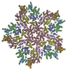
| Supplemental images |
|---|
- Downloads & links
-EMDB archive
| Map data | emd_16698.map.gz | 25.1 MB |  EMDB map data format EMDB map data format | |
|---|---|---|---|---|
| Header (meta data) | emd-16698-v30.xmlemd-16698.xml | 17.3 KB 17.3 KB | Display Display | EMDB header |
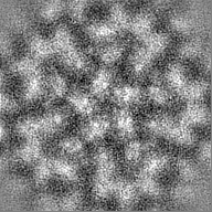
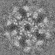
| Images |  emd_16698.png emd_16698.png | 134.7 KB | ||
| Others | emd_16698_half_map_1.map.gzemd_16698_half_map_2.map.gz | 25.1 MB 25.1 MB | ||
| Archive directory |  http://ftp.pdbj.org/pub/emdb/structures/EMD-16698ftp://ftp.pdbj.org/pub/emdb/structures/EMD-16698 http://ftp.pdbj.org/pub/emdb/structures/EMD-16698ftp://ftp.pdbj.org/pub/emdb/structures/EMD-16698 | HTTPS FTP |
-Related structure data


-Links
| EMDB pages | EMDB (EBI/PDBe) / EMDataResource |
|---|
-Map
| File | Download / File: emd_16698.map.gz / Format: CCP4 / Size: 27 MB / Type: IMAGE STORED AS FLOATING POINT NUMBER (4 BYTES) | ||||||||||||||||||||||||||||||||||||
|---|---|---|---|---|---|---|---|---|---|---|---|---|---|---|---|---|---|---|---|---|---|---|---|---|---|---|---|---|---|---|---|---|---|---|---|---|---|
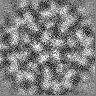
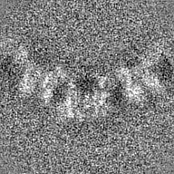
| Annotation | Subtomogram average of HIV-1 CA protein pentamer from capsid-like particles assembled with inositol hexakisphosphate | ||||||||||||||||||||||||||||||||||||
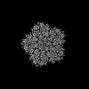
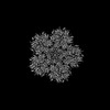
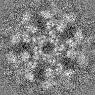
| Projections & slices | Image control
Images are generated by Spider. | ||||||||||||||||||||||||||||||||||||
| Voxel size | X=Y=Z: 1.379 Å | ||||||||||||||||||||||||||||||||||||
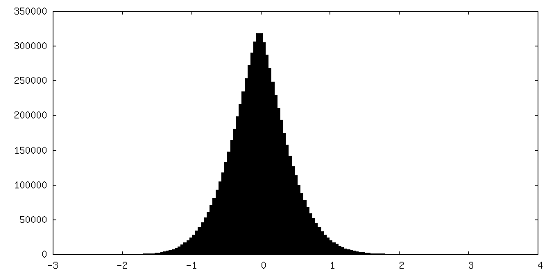
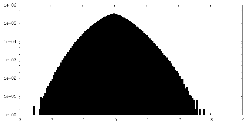
| Density |
| ||||||||||||||||||||||||||||||||||||
| Symmetry | Space group: 1 | ||||||||||||||||||||||||||||||||||||
| Details | EMDB XML:
|
 Z (Sec.)
 Y (Row.)
 X (Col.)


-Supplemental data
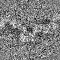
-Half map: Subtomogram average of HIV-1 CA protein pentamer from...
| File | emd_16698_half_map_1.map | ||||||||||||
|---|---|---|---|---|---|---|---|---|---|---|---|---|---|
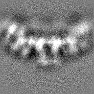
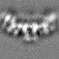
| Annotation | Subtomogram average of HIV-1 CA protein pentamer from capsid-like particles assembled with inositol hexakisphosphate, half map A | ||||||||||||
| Projections & Slices |
| ||||||||||||
| Density Histograms |
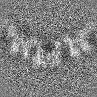

-Half map: Subtomogram average of HIV-1 CA protein pentamer from...
| File | emd_16698_half_map_2.map | ||||||||||||
|---|---|---|---|---|---|---|---|---|---|---|---|---|---|
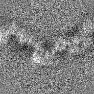
| Annotation | Subtomogram average of HIV-1 CA protein pentamer from capsid-like particles assembled with inositol hexakisphosphate, half map B | ||||||||||||
| Projections & Slices |
| ||||||||||||
| Density Histograms |

- Sample components
-Entire : HIV-1 CA protein capsid-like particles assembled in the presence ...
| Entire | Name: HIV-1 CA protein capsid-like particles assembled in the presence of inositol hexakisphosphate |
|---|---|
| Components |
|
-Supramolecule #1: HIV-1 CA protein capsid-like particles assembled in the presence ...
| Supramolecule | Name: HIV-1 CA protein capsid-like particles assembled in the presence of inositol hexakisphosphate type: complex / ID: 1 / Chimera: Yes / Parent: 0 |
|---|---|
| Source (natural) | Organism: |
-Experimental details
-Structure determination
| Method | cryo EM |
|---|---|
 Processing Processing | subtomogram averaging |
| Aggregation state | particle |
-Sample preparation
| Buffer | pH: 6.2 Component:
| ||||||||||||
|---|---|---|---|---|---|---|---|---|---|---|---|---|---|
| Grid | Model: C-flat-2/2 / Material: COPPER / Mesh: 300 / Pretreatment - Type: GLOW DISCHARGE / Pretreatment - Time: 30 sec. / Pretreatment - Atmosphere: AIR Details: The grid was glow discharged for 30 seconds at a current of 20 mA. | ||||||||||||
| Vitrification | Cryogen name: ETHANE / Chamber humidity: 100 % / Chamber temperature: 277.15 K / Instrument: FEI VITROBOT MARK IV |
- Electron microscopy
| Microscope | FEI TITAN KRIOS |
|---|---|
| Specialist optics | Energy filter - Name: GIF Bioquantum / Energy filter - Slit width: 20 eV |
| Image recording | Film or detector model: GATAN K2 SUMMIT (4k x 4k) / Detector mode: COUNTING / Average electron dose: 3.0 e/Å2 |
| Electron beam | Acceleration voltage: 300 kV / Electron source:  FIELD EMISSION GUN FIELD EMISSION GUN |
| Electron optics | Illumination mode: FLOOD BEAM / Imaging mode: BRIGHT FIELD / Cs: 2.7 mm / Nominal defocus max: 4.5 µm / Nominal defocus min: 1.5 µm / Nominal magnification: 105000 |
| Sample stage | Specimen holder model: FEI TITAN KRIOS AUTOGRID HOLDER / Cooling holder cryogen: NITROGEN |
| Experimental equipment |  Model: Titan Krios / Image courtesy: FEI Company |
-Image processing
| Final reconstruction | Applied symmetry - Point group: C5 (5 fold cyclic) / Algorithm: BACK PROJECTION / Resolution.type: BY AUTHOR / Resolution: 6.2 Å / Resolution method: FSC 0.143 CUT-OFF / Software - Name: subTOM / Number subtomograms used: 5244 |
|---|---|
| Extraction | Number tomograms: 66 / Number images used: 5264 / Software - Name: MATLAB Details: Pentamer positions in HIV-1 CA protein capsid-like particles were identified by identifying patterns of 5 aligned hexamer positions corresponding to pentamer geometry using MATLAB. |
| Final angle assignment | Type: OTHER / Software - Name: subTOM |
