 Movie
Movie Controller

[English] 日本語
 Yorodumi
- PDB-8ejl: Structure of HIV-1 capsid declination in complex with CPSF6-FG peptide -
+ Open data

- Basic information
| Entry | Database: PDB / ID: 8ejl | |||||||||
|---|---|---|---|---|---|---|---|---|---|---|
| Title | Structure of HIV-1 capsid declination in complex with CPSF6-FG peptide | |||||||||
 Components Components |
| |||||||||
 Keywords Keywords | VIRAL PROTEIN / HIV-1 / capsid / declination / pentamer / hexamer | |||||||||
| Function / homology |  Function and homology information Function and homology informationexon-exon junction complex binding / mRNA alternative polyadenylation / positive regulation of RNA export from nucleus / mRNA cleavage factor complex / co-transcriptional mRNA 3'-end processing, cleavage and polyadenylation pathway / interchromatin granule / perichromatin fibrils / Processing of Intronless Pre-mRNAs / mRNA cleavage and polyadenylation specificity factor complex / mRNA 3'-end processing ...exon-exon junction complex binding / mRNA alternative polyadenylation / positive regulation of RNA export from nucleus / mRNA cleavage factor complex / co-transcriptional mRNA 3'-end processing, cleavage and polyadenylation pathway / interchromatin granule / perichromatin fibrils / Processing of Intronless Pre-mRNAs / mRNA cleavage and polyadenylation specificity factor complex / mRNA 3'-end processing / Signaling by cytosolic FGFR1 fusion mutants / paraspeckles / mRNA 3'-end processing / RNA Polymerase II Transcription Termination / protein heterotetramerization / viral budding via host ESCRT complex / ribosomal large subunit binding / Processing of Capped Intron-Containing Pre-mRNA / Signaling by FGFR1 in disease / protein tetramerization / host multivesicular body / ISG15 antiviral mechanism / mRNA processing / viral nucleocapsid / nuclear speck / ribonucleoprotein complex / viral translational frameshifting / mRNA binding / host cell nucleus / host cell plasma membrane / virion membrane / structural molecule activity / RNA binding / zinc ion binding / nucleoplasm / membrane / nucleus / cytoplasm Similarity search - Function | |||||||||
| Biological species |   Human immunodeficiency virus 1 Human immunodeficiency virus 1 Homo sapiens (human) Homo sapiens (human) | |||||||||
| Method | ELECTRON MICROSCOPY / single particle reconstruction / cryo EM / Resolution: 3.9 Å | |||||||||
 Authors Authors | Pornillos, O. / Ganser-Pornillos, B.K. / Schirra, R.T. / dos Santos, N.F.B. | |||||||||
| Funding support |  United States, 2items United States, 2items
| |||||||||
 Citation Citation | Journal: Nat Struct Mol Biol / Year: 2023 Title: A molecular switch modulates assembly and host factor binding of the HIV-1 capsid. Authors: Randall T Schirra / Nayara F B Dos Santos / Kaneil K Zadrozny / Iga Kucharska / Barbie K Ganser-Pornillos / Owen Pornillos /  Abstract: The HIV-1 capsid is a fullerene cone made of quasi-equivalent hexamers and pentamers of the viral CA protein. Typically, quasi-equivalent assembly of viral capsid subunits is controlled by a ...The HIV-1 capsid is a fullerene cone made of quasi-equivalent hexamers and pentamers of the viral CA protein. Typically, quasi-equivalent assembly of viral capsid subunits is controlled by a molecular switch. Here, we identify a Thr-Val-Gly-Gly motif that modulates CA hexamer/pentamer switching by folding into a 3 helix in the pentamer and random coil in the hexamer. Manipulating the coil/helix configuration of the motif allowed us to control pentamer and hexamer formation in a predictable manner, thus proving its function as a molecular switch. Importantly, the switch also remodels the common binding site for host factors that are critical for viral replication and the new ultra-potent HIV-1 inhibitor lenacapavir. This study reveals that a critical assembly element also modulates the post-assembly and viral replication functions of the HIV-1 capsid and provides new insights on capsid function and inhibition. | |||||||||
| History |
|
- Structure visualization
| Structure viewer | Molecule: MolmilJmol/JSmol |
|---|
- Downloads & links
-Download
| PDBx/mmCIF format | 8ejl.cif.gz | 116 KB | Display | PDBx/mmCIF format |
|---|---|---|---|---|
| PDB format | pdb8ejl.ent.gz | 86.1 KB | Display | PDB format |
| PDBx/mmJSON format | 8ejl.json.gz | Tree view | PDBx/mmJSON format | |
| Others |  Other downloads Other downloads |
-Validation report
| Arichive directory | https://data.pdbj.org/pub/pdb/validation_reports/ej/8ejlftp://data.pdbj.org/pub/pdb/validation_reports/ej/8ejl | HTTPS FTP |
|---|
-Related structure data
| Related structure data |  28186MC  7urnC 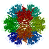 7urtC  8eepC 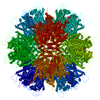 8eetC C: citing same article ( M: map data used to model this data |
|---|---|
| Similar structure data |


-Links
 PDBj
PDBj


- Assembly
| Deposited unit | 
|
|---|---|
| 1 | x 5
|
| 2 |
|
| 3 | 
|
| Symmetry | Point symmetry: (Schoenflies symbol: C5 (5 fold cyclic)) |
-Components
| #1: Protein | Mass: 25630.426 Da / Num. of mol.: 4 Source method: isolated from a genetically manipulated source Source: (gene. exp.) Human immunodeficiency virus 1 / Strain: NL4-3 / Gene: Gag-pol / Production host:  #2: Protein/peptide | Mass: 1708.952 Da / Num. of mol.: 2 / Fragment: UNP residues 313-327 / Source method: obtained synthetically / Source: (synth.) Homo sapiens (human) / References: UniProt: Q16630 |
|---|
-Experimental details
-Experiment
| Experiment | Method: ELECTRON MICROSCOPY |
|---|---|
| EM experiment | Aggregation state: PARTICLE / 3D reconstruction method: single particle reconstruction |
- Sample preparation
| Component | Name: Capsid-like particles of in vitro assembled HIV-1 CA protein in complex with CPSF6-FG peptide Type: COMPLEX Details: Capsids (0.27 mM CA) were incubated with peptide (3 mM) for 1 hour in ice Entity ID: all / Source: MULTIPLE SOURCES |
|---|---|
| Source (natural) | Organism: Human immunodeficiency virus 1 |
| Source (recombinant) | Organism: |
| Details of virus | Empty: YES / Enveloped: NO / Isolate: OTHER / Type: VIRUS-LIKE PARTICLE |
| Buffer solution | pH: 6 |
| Specimen | Embedding applied: NO / Shadowing applied: NO / Staining applied: NO / Vitrification applied: YES |
| Vitrification | Instrument: HOMEMADE PLUNGER / Cryogen name: ETHANE / Details: Manual plunge-freezing |
- Electron microscopy imaging
| Experimental equipment |  Model: Titan Krios / Image courtesy: FEI Company |
|---|---|
| Microscopy | Model: FEI TITAN KRIOS |
| Electron gun | Electron source:  FIELD EMISSION GUN / Accelerating voltage: 300 kV / Illumination mode: FLOOD BEAM FIELD EMISSION GUN / Accelerating voltage: 300 kV / Illumination mode: FLOOD BEAM |
| Electron lens | Mode: BRIGHT FIELD / Nominal defocus max: 2500 nm / Nominal defocus min: 500 nm |
| Image recording | Electron dose: 50 e/Å2 / Film or detector model: GATAN K3 (6k x 4k) |
- Processing
| Software | Name: PHENIX / Version: 1.20.1_4487: / Classification: refinement | ||||||||||||||||||||||||||||||||||||
|---|---|---|---|---|---|---|---|---|---|---|---|---|---|---|---|---|---|---|---|---|---|---|---|---|---|---|---|---|---|---|---|---|---|---|---|---|---|
| EM software |
| ||||||||||||||||||||||||||||||||||||
| CTF correction | Type: PHASE FLIPPING AND AMPLITUDE CORRECTION | ||||||||||||||||||||||||||||||||||||
| 3D reconstruction | Resolution: 3.9 Å / Resolution method: FSC 0.143 CUT-OFF / Num. of particles: 166463 / Symmetry type: POINT | ||||||||||||||||||||||||||||||||||||
| Atomic model building | Protocol: OTHER / Space: REAL | ||||||||||||||||||||||||||||||||||||
| Atomic model building | 3D fitting-ID: 1 / Source name: PDB / Type: experimental model
| ||||||||||||||||||||||||||||||||||||
| Refine LS restraints |
|

