 ムービー
ムービー コントローラー


+ データを開く

- 基本情報
基本情報
| 登録情報 | データベース: PDB / ID: 8efe | ||||||
|---|---|---|---|---|---|---|---|
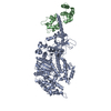
| タイトル | Human beta-cardiac myosin II bound to ADP-MG2+ and the associated essential light chain | ||||||
 要素 要素 |
| ||||||
 キーワード キーワード | CONTRACTILE PROTEIN / actin / tropomyosin / myosin / cardiac | ||||||
| 機能・相同性 |  機能・相同性情報 機能・相同性情報regulation of slow-twitch skeletal muscle fiber contraction / regulation of the force of skeletal muscle contraction / Striated Muscle Contraction / muscle myosin complex / regulation of the force of heart contraction / transition between fast and slow fiber / myosin filament / adult heart development / muscle filament sliding / cardiac muscle hypertrophy in response to stress ...regulation of slow-twitch skeletal muscle fiber contraction / regulation of the force of skeletal muscle contraction / Striated Muscle Contraction / muscle myosin complex / regulation of the force of heart contraction / transition between fast and slow fiber / myosin filament / adult heart development / muscle filament sliding / cardiac muscle hypertrophy in response to stress / myosin complex / myosin II complex / structural constituent of muscle / ventricular cardiac muscle tissue morphogenesis / microfilament motor activity / myofibril / skeletal muscle contraction / striated muscle contraction / ATP metabolic process / cardiac muscle contraction / stress fiber / regulation of heart rate / muscle contraction / sarcomere / Z disc / actin filament binding / calmodulin binding / calcium ion binding / ATP binding / cytoplasm 類似検索 - 分子機能 | ||||||
| 生物種 |  Homo sapiens (ヒト) Homo sapiens (ヒト) | ||||||
| 手法 | 電子顕微鏡法 / 単粒子再構成法 / クライオ電子顕微鏡法 / 解像度: 3.8 Å | ||||||
 データ登録者 データ登録者 | Doran, M.H. / Lehman, W. / Rynkiewicz, M.J. | ||||||
| 資金援助 |  米国, 1件 米国, 1件
| ||||||
 引用 引用 | ジャーナル: J Gen Physiol / 年: 2023 タイトル: Conformational changes linked to ADP release from human cardiac myosin bound to actin-tropomyosin. 著者: Matthew H Doran / Michael J Rynkiewicz / David Rasicci / Skylar M L Bodt / Meaghan E Barry / Esther Bullitt / Christopher M Yengo / Jeffrey R Moore / William Lehman / 要旨: Following binding to the thin filament, β-cardiac myosin couples ATP-hydrolysis to conformational rearrangements in the myosin motor that drive myofilament sliding and cardiac ventricular ...Following binding to the thin filament, β-cardiac myosin couples ATP-hydrolysis to conformational rearrangements in the myosin motor that drive myofilament sliding and cardiac ventricular contraction. However, key features of the cardiac-specific actin-myosin interaction remain uncertain, including the structural effect of ADP release from myosin, which is rate-limiting during force generation. In fact, ADP release slows under experimental load or in the intact heart due to the afterload, thereby adjusting cardiac muscle power output to meet physiological demands. To further elucidate the structural basis of this fundamental process, we used a combination of cryo-EM reconstruction methodologies to determine structures of the human cardiac actin-myosin-tropomyosin filament complex at better than 3.4 Å-resolution in the presence and in the absence of Mg2+·ADP. Focused refinements of the myosin motor head and its essential light chains in these reconstructions reveal that small changes in the nucleotide-binding site are coupled to significant rigid body movements of the myosin converter domain and a 16-degree lever arm swing. Our structures provide a mechanistic framework to understand the effect of ADP binding and release on human cardiac β-myosin, and offer insights into the force-sensing mechanism displayed by the cardiac myosin motor. | ||||||
| 履歴 |
|
- 構造の表示
構造の表示
| 構造ビューア | 分子: MolmilJmol/JSmol |
|---|
- ダウンロードとリンク
-ダウンロード
| PDBx/mmCIF形式 | 8efe.cif.gz | 173.3 KB | 表示 | PDBx/mmCIF形式 |
|---|---|---|---|---|
| PDB形式 | pdb8efe.ent.gz | 132.5 KB | 表示 | PDB形式 |
| PDBx/mmJSON形式 | 8efe.json.gz | ツリー表示 | PDBx/mmJSON形式 | |
| その他 |  その他のダウンロード その他のダウンロード |
-検証レポート
| アーカイブディレクトリ | https://data.pdbj.org/pub/pdb/validation_reports/ef/8efeftp://data.pdbj.org/pub/pdb/validation_reports/ef/8efe | HTTPS FTP |
|---|
-関連構造データ
-リンク
 PDBj
PDBj


- 集合体
集合体
| 登録構造単位 | 
|
|---|---|
| 1 |
|
-要素
| #1: タンパク質 | 分子量: 99157.695 Da / 分子数: 1 / 由来タイプ: 組換発現 / 由来: (組換発現) Homo sapiens (ヒト) / 遺伝子: MYH7, MYHCB / 細胞株 (発現宿主): C2C12 / 発現宿主: |
|---|---|
| #2: タンパク質 | 分子量: 20620.490 Da / 分子数: 1 / 由来タイプ: 天然 詳細: Mouse skeletal muscle essential light chain 1/3 that was purified with the human cardiac myosin II. 由来: (天然) |
| #3: 化合物 | ChemComp-MG /   分子量: 24.305 Da / 分子数: 1 / 由来タイプ: 合成 / 式: Mg / タイプ: SUBJECT OF INVESTIGATION 分子量: 24.305 Da / 分子数: 1 / 由来タイプ: 合成 / 式: Mg / タイプ: SUBJECT OF INVESTIGATION |
| #4: 化合物 | ChemComp-ADP /   分子量: 427.201 Da / 分子数: 1 / 由来タイプ: 合成 / 式: C10H15N5O10P2 / タイプ: SUBJECT OF INVESTIGATION / コメント: ADP, エネルギー貯蔵分子*YM 分子量: 427.201 Da / 分子数: 1 / 由来タイプ: 合成 / 式: C10H15N5O10P2 / タイプ: SUBJECT OF INVESTIGATION / コメント: ADP, エネルギー貯蔵分子*YM |
| 研究の焦点であるリガンドがあるか | Y |
-実験情報
-実験
| 実験 | 手法: 電子顕微鏡法 |
|---|---|
| EM実験 | 試料の集合状態: FILAMENT / 3次元再構成法: 単粒子再構成法 |
- 試料調製
試料調製
| 構成要素 |
| ||||||||||||||||||||||||
|---|---|---|---|---|---|---|---|---|---|---|---|---|---|---|---|---|---|---|---|---|---|---|---|---|---|
| 分子量 | 実験値: NO | ||||||||||||||||||||||||
| 由来(天然) |
| ||||||||||||||||||||||||
| 由来(組換発現) | 生物種: | ||||||||||||||||||||||||
| 緩衝液 | pH: 7 | ||||||||||||||||||||||||
| 試料 | 濃度: 0.13 mg/ml / 包埋: NO / シャドウイング: NO / 染色: NO / 凍結: YES 詳細: This complex was part of a larger filament containing actin-tropomyosin. The map was a result of a focused single particle approach. | ||||||||||||||||||||||||
| 試料支持 | 詳細: 15 mA was used in the Pelco Easiglow glow discharge machine グリッドの材料: GOLD / グリッドのサイズ: 400 divisions/in. / グリッドのタイプ: Quantifoil R1.2/1.3 | ||||||||||||||||||||||||
| 急速凍結 | 装置: FEI VITROBOT MARK III / 凍結剤: ETHANE / 湿度: 100 % / 凍結前の試料温度: 283 K |
- 電子顕微鏡撮影
| 実験機器 |  モデル: Titan Krios / 画像提供: FEI Company |
|---|---|
| 顕微鏡 | モデル: FEI TITAN KRIOS |
| 電子銃 | 電子線源:  FIELD EMISSION GUN / 加速電圧: 300 kV / 照射モード: SPOT SCAN FIELD EMISSION GUN / 加速電圧: 300 kV / 照射モード: SPOT SCAN |
| 電子レンズ | モード: BRIGHT FIELD / 倍率(補正後): 80000 X / 最大 デフォーカス(公称値): 3000 nm / 最小 デフォーカス(公称値): 700 nm |
| 試料ホルダ | 凍結剤: NITROGEN 試料ホルダーモデル: FEI TITAN KRIOS AUTOGRID HOLDER |
| 撮影 | 平均露光時間: 3.12 sec. / 電子線照射量: 53.7 e/Å2 / 検出モード: COUNTING / フィルム・検出器のモデル: GATAN K3 (6k x 4k) / 撮影したグリッド数: 4 / 実像数: 3961 |
| 電子光学装置 | エネルギーフィルター名称: In-column Omega Filter エネルギーフィルタースリット幅: 20 eV |
| 画像スキャン | 動画フレーム数/画像: 35 / 利用したフレーム数/画像: 1-35 |
- 解析
解析
| ソフトウェア | 名称: PHENIX / バージョン: 1.19.1_4122: / 分類: 精密化 | ||||||||||||||||||||||||||||||||||||||||
|---|---|---|---|---|---|---|---|---|---|---|---|---|---|---|---|---|---|---|---|---|---|---|---|---|---|---|---|---|---|---|---|---|---|---|---|---|---|---|---|---|---|
| EMソフトウェア |
| ||||||||||||||||||||||||||||||||||||||||
| CTF補正 | タイプ: PHASE FLIPPING AND AMPLITUDE CORRECTION | ||||||||||||||||||||||||||||||||||||||||
| 粒子像の選択 | 選択した粒子像数: 131194 | ||||||||||||||||||||||||||||||||||||||||
| 対称性 | 点対称性: C1 (非対称) | ||||||||||||||||||||||||||||||||||||||||
| 3次元再構成 | 解像度: 3.8 Å / 解像度の算出法: FSC 0.143 CUT-OFF / 粒子像の数: 68358 / 対称性のタイプ: POINT | ||||||||||||||||||||||||||||||||||||||||
| 原子モデル構築 | プロトコル: OTHER / 空間: REAL | ||||||||||||||||||||||||||||||||||||||||
| 原子モデル構築 | PDB-ID: 6X5Z Accession code: 6X5Z / Source name: PDB / タイプ: experimental model | ||||||||||||||||||||||||||||||||||||||||
| 拘束条件 |
|

