 Movie
Movie Controller


+ Open data

- Basic information
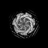
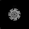
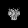
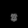
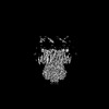
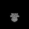
| Entry | Database: PDB / ID: 7uw5 | ||||||
|---|---|---|---|---|---|---|---|
| Title | EcMscK G924S mutant in a closed conformation | ||||||
 Components Components | Mechanosensitive channel MscK | ||||||
 Keywords Keywords | TRANSPORT PROTEIN / MEMBRANE PROTEIN / MECHANOSENSATION / ION CHANNEL | ||||||
| Function / homology |  Function and homology information Function and homology informationintracellular water homeostasis / response to potassium ion / mechanosensitive monoatomic ion channel activity / potassium ion transport / plasma membrane Similarity search - Function | ||||||
| Biological species |  | ||||||
| Method | ELECTRON MICROSCOPY / single particle reconstruction / cryo EM / Resolution: 3.84 Å | ||||||
 Authors Authors | Mount, J.W. / Yuan, P. | ||||||
| Funding support |  United States, 1items United States, 1items
| ||||||
 Citation Citation | Journal: Nat Commun / Year: 2022 Title: Structural basis for mechanotransduction in a potassium-dependent mechanosensitive ion channel. Authors: Jonathan Mount / Grigory Maksaev / Brock T Summers / James A J Fitzpatrick / Peng Yuan / Abstract: Mechanosensitive channels of small conductance, found in many living organisms, open under elevated membrane tension and thus play crucial roles in biological response to mechanical stress. Amongst ...Mechanosensitive channels of small conductance, found in many living organisms, open under elevated membrane tension and thus play crucial roles in biological response to mechanical stress. Amongst these channels, MscK is unique in that its activation also requires external potassium ions. To better understand this dual gating mechanism by force and ligand, we elucidate distinct structures of MscK along the gating cycle using cryo-electron microscopy. The heptameric channel comprises three layers: a cytoplasmic domain, a periplasmic gating ring, and a markedly curved transmembrane domain that flattens and expands upon channel opening, which is accompanied by dilation of the periplasmic ring. Furthermore, our results support a potentially unifying mechanotransduction mechanism in ion channels depicted as flattening and expansion of the transmembrane domain. | ||||||
| History |
|
- Structure visualization
| Structure viewer | Molecule: MolmilJmol/JSmol |
|---|
- Downloads & links
-Download
| PDBx/mmCIF format | 7uw5.cif.gz | 815.3 KB | Display | PDBx/mmCIF format |
|---|---|---|---|---|
| PDB format | pdb7uw5.ent.gz | 600.9 KB | Display | PDB format |
| PDBx/mmJSON format | 7uw5.json.gz | Tree view | PDBx/mmJSON format | |
| Others |  Other downloads Other downloads |
-Validation report
| Arichive directory | https://data.pdbj.org/pub/pdb/validation_reports/uw/7uw5ftp://data.pdbj.org/pub/pdb/validation_reports/uw/7uw5 | HTTPS FTP |
|---|
-Related structure data
| Related structure data | 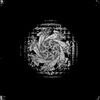 26823MC 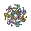 7ux1C M: map data used to model this data C: citing same article ( |
|---|---|
| Similar structure data |
-Links
 PDBj
PDBj


- Assembly
| Deposited unit | 
|
|---|---|
| 1 |
|
-Components
| #1: Protein | Mass: 127378.789 Da / Num. of mol.: 7 / Mutation: G924S Source method: isolated from a genetically manipulated source Source: (gene. exp.)  Komagataella pastoris (fungus) / References: UniProt: P77338 Komagataella pastoris (fungus) / References: UniProt: P77338 |
|---|
-Experimental details
-Experiment
| Experiment | Method: ELECTRON MICROSCOPY |
|---|---|
| EM experiment | Aggregation state: PARTICLE / 3D reconstruction method: single particle reconstruction |
- Sample preparation
| Component | Name: E.coli MscK / Type: COMPLEX / Details: Homomeric Heptamer / Entity ID: all / Source: RECOMBINANT | |||||||||||||||||||||||||
|---|---|---|---|---|---|---|---|---|---|---|---|---|---|---|---|---|---|---|---|---|---|---|---|---|---|---|
| Molecular weight | Experimental value: NO | |||||||||||||||||||||||||
| Source (natural) | Organism: | |||||||||||||||||||||||||
| Source (recombinant) | Organism: Komagataella pastoris (fungus) / Plasmid: PV-1 | |||||||||||||||||||||||||
| Buffer solution | pH: 8 Details: Micrographs were pooled from protein purified in the presence of either 150mM NaCl or 150mM KCl, respectively. Buffers were prepared fresh, degassed, and filtered through a 0.45 um Durapore PVDF membrane | |||||||||||||||||||||||||
| Buffer component |
| |||||||||||||||||||||||||
| Specimen | Conc.: 7 mg/ml / Embedding applied: NO / Shadowing applied: NO / Staining applied: NO / Vitrification applied: YES / Details: The specimen was homogeneous and monodisperse. | |||||||||||||||||||||||||
| Specimen support | Grid material: COPPER / Grid mesh size: 300 divisions/in. / Grid type: Quantifoil R2/2 | |||||||||||||||||||||||||
| Vitrification | Instrument: FEI VITROBOT MARK IV / Cryogen name: ETHANE / Humidity: 100 % / Chamber temperature: 277.15 K |
- Electron microscopy imaging
| Microscopy | Model: TFS GLACIOS |
|---|---|
| Electron gun | Electron source:  FIELD EMISSION GUN / Accelerating voltage: 200 kV / Illumination mode: FLOOD BEAM FIELD EMISSION GUN / Accelerating voltage: 200 kV / Illumination mode: FLOOD BEAM |
| Electron lens | Mode: BRIGHT FIELD / Nominal magnification: 150000 X / Nominal defocus max: 2400 nm / Nominal defocus min: 600 nm / Alignment procedure: BASIC |
| Specimen holder | Cryogen: NITROGEN |
| Image recording | Electron dose: 46.16 e/Å2 / Detector mode: COUNTING / Film or detector model: FEI FALCON IV (4k x 4k) |
- Processing
| EM software | Name: cryoSPARC / Version: 3.1 / Category: 3D reconstruction |
|---|---|
| CTF correction | Type: NONE |
| Symmetry | Point symmetry: C7 (7 fold cyclic) |
| 3D reconstruction | Resolution: 3.84 Å / Resolution method: FSC 0.143 CUT-OFF / Num. of particles: 134427 / Num. of class averages: 1 / Symmetry type: POINT |
| Atomic model building | Protocol: AB INITIO MODEL / Space: REAL |
| Atomic model building | PDB-ID: 7UW5 Pdb chain-ID: A / Accession code: 7UW5 / Pdb chain residue range: 496-1079 / Source name: PDB / Type: experimental model |
