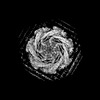
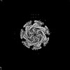
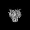
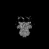
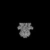
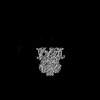
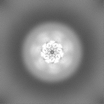
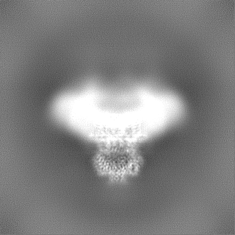
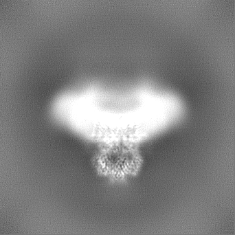
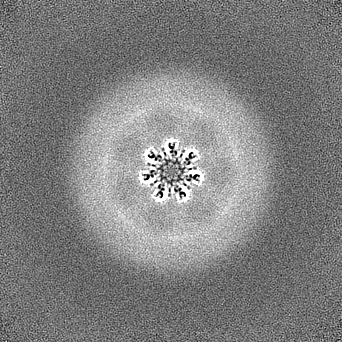
- EMDB-26872: Locally refined core of EcMscK in a closed conformation -
+
Open data
ID or keywords:
Loading...
-
Basic information
Entry
Database: EMDB / ID: EMD-26872
Title
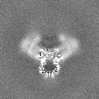
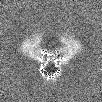
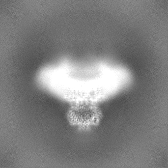
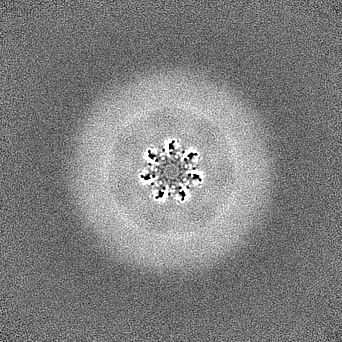
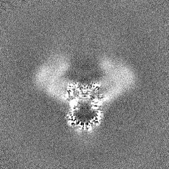
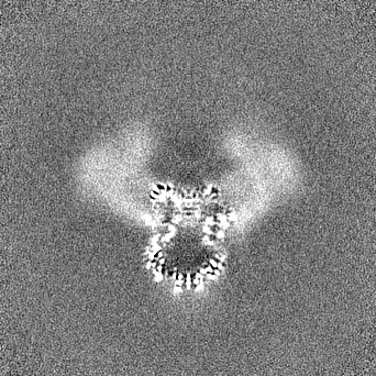
Locally refined core of EcMscK in a closed conformation
Map data
Local Refinement of EcMscK from the CTD through TM 9.
Sample
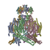
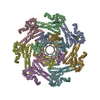
Complex: EcMscK
Protein or peptide: EcMscK
Keywords
MEMBRANE PROTEIN / MECHANOSENSATION / ION CHANNEL / TRANSPORT PROTEIN
Function / homology
Function and homology information
intracellular water homeostasis / response to potassium ion / mechanosensitive monoatomic ion channel activity / potassium ion transport / plasma membrane Similarity search - Function
Mechanosensitive ion channel MscS, porin domain / Mechanosensitive ion channel inner membrane domain 1 / : / Mechanosensitive ion channel inner membrane domain 1 / Mechanosensitive ion channel porin domain / : / : / Mechanosensitive ion channel MscS, C-terminal / Mechanosensitive ion channel, transmembrane helices 2/3 / Mechanosensitive ion channel MscS, conserved site ...Mechanosensitive ion channel MscS, porin domain / Mechanosensitive ion channel inner membrane domain 1 / : / Mechanosensitive ion channel inner membrane domain 1 / Mechanosensitive ion channel porin domain / : / : / Mechanosensitive ion channel MscS, C-terminal / Mechanosensitive ion channel, transmembrane helices 2/3 / Mechanosensitive ion channel MscS, conserved site / Uncharacterized protein family UPF0003 signature. / Mechanosensitive ion channel MscS, C-terminal / Mechanosensitive ion channel MscS, transmembrane-2 / Mechanosensitive ion channel MscS / Mechanosensitive ion channel, beta-domain / Mechanosensitive ion channel MscS, beta-domain superfamily / LSM domain superfamily / P-type ATPase, transmembrane domain superfamily Similarity search - Domain/homology
National Institutes of Health/National Institute of Neurological Disorders and Stroke (NIH/NINDS)
NS099341
United States
Citation
Journal: Nat Commun / Year: 2022 Title: Structural basis for mechanotransduction in a potassium-dependent mechanosensitive ion channel. Authors: Jonathan Mount / Grigory Maksaev / Brock T Summers / James A J Fitzpatrick / Peng Yuan / Abstract: Mechanosensitive channels of small conductance, found in many living organisms, open under elevated membrane tension and thus play crucial roles in biological response to mechanical stress. Amongst ...Mechanosensitive channels of small conductance, found in many living organisms, open under elevated membrane tension and thus play crucial roles in biological response to mechanical stress. Amongst these channels, MscK is unique in that its activation also requires external potassium ions. To better understand this dual gating mechanism by force and ligand, we elucidate distinct structures of MscK along the gating cycle using cryo-electron microscopy. The heptameric channel comprises three layers: a cytoplasmic domain, a periplasmic gating ring, and a markedly curved transmembrane domain that flattens and expands upon channel opening, which is accompanied by dilation of the periplasmic ring. Furthermore, our results support a potentially unifying mechanotransduction mechanism in ion channels depicted as flattening and expansion of the transmembrane domain.
In the structure databanks used in Yorodumi, some data are registered as the other names, "COVID-19 virus" and "2019-nCoV". Here are the details of the virus and the list of structure data.
Jan 31, 2019. EMDB accession codes are about to change! (news from PDBe EMDB page)
EMDB accession codes are about to change! (news from PDBe EMDB page)
The allocation of 4 digits for EMDB accession codes will soon come to an end. Whilst these codes will remain in use, new EMDB accession codes will include an additional digit and will expand incrementally as the available range of codes is exhausted. The current 4-digit format prefixed with “EMD-” (i.e. EMD-XXXX) will advance to a 5-digit format (i.e. EMD-XXXXX), and so on. It is currently estimated that the 4-digit codes will be depleted around Spring 2019, at which point the 5-digit format will come into force.
The EM Navigator/Yorodumi systems omit the EMD- prefix.
Related info.:Q: What is EMD? / ID/Accession-code notation in Yorodumi/EM Navigator
Yorodumi is a browser for structure data from EMDB, PDB, SASBDB, etc.
This page is also the successor to EM Navigator detail page, and also detail information page/front-end page for Omokage search.
The word "yorodu" (or yorozu) is an old Japanese word meaning "ten thousand". "mi" (miru) is to see.
Related info.:EMDB / PDB / SASBDB / Comparison of 3 databanks / Yorodumi Search / Aug 31, 2016. New EM Navigator & Yorodumi / Yorodumi Papers / Jmol/JSmol / Function and homology information / Changes in new EM Navigator and Yorodumi
 Movie
Movie Controller


 Open data
 Basic information
 Map data
 Sample
 Keywords
 Function and homology information
 Authors
 United States, 1 items
  Citation
 Structure visualization
 Downloads & links
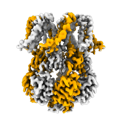 emd_26872.png
 http://ftp.pdbj.org/pub/emdb/structures/EMD-26872


 Z (Sec.)
 Y (Row.)
 X (Col.)


 Sample components
 Komagataella pastoris (fungus)
 Processing
 Electron microscopy
 FIELD EMISSION GUN