 Movie
Movie Controller

[English] 日本語
 Yorodumi
- EMDB-26876: Locally refined core of EcMscK G924S in an intermediate conformation -
+ Open data

- Basic information
| Entry | 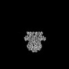 | |||||||||
|---|---|---|---|---|---|---|---|---|---|---|
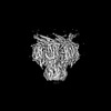
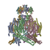
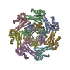
| Title | Locally refined core of EcMscK G924S in an intermediate conformation | |||||||||
 Map data Map data | Locally refined core of EcMscK G924S in an intermediate conformation | |||||||||
 Sample Sample |
| |||||||||
| Biological species |  | |||||||||
| Method | single particle reconstruction / cryo EM / Resolution: 3.85 Å | |||||||||
 Authors Authors | Mount JW / Yuan P | |||||||||
| Funding support |  United States, 1 items United States, 1 items
| |||||||||
 Citation Citation | Journal: Nat Commun / Year: 2022 Title: Structural basis for mechanotransduction in a potassium-dependent mechanosensitive ion channel. Authors: Jonathan Mount / Grigory Maksaev / Brock T Summers / James A J Fitzpatrick / Peng Yuan / Abstract: Mechanosensitive channels of small conductance, found in many living organisms, open under elevated membrane tension and thus play crucial roles in biological response to mechanical stress. Amongst ...Mechanosensitive channels of small conductance, found in many living organisms, open under elevated membrane tension and thus play crucial roles in biological response to mechanical stress. Amongst these channels, MscK is unique in that its activation also requires external potassium ions. To better understand this dual gating mechanism by force and ligand, we elucidate distinct structures of MscK along the gating cycle using cryo-electron microscopy. The heptameric channel comprises three layers: a cytoplasmic domain, a periplasmic gating ring, and a markedly curved transmembrane domain that flattens and expands upon channel opening, which is accompanied by dilation of the periplasmic ring. Furthermore, our results support a potentially unifying mechanotransduction mechanism in ion channels depicted as flattening and expansion of the transmembrane domain. | |||||||||
| History |
|
- Structure visualization
| Supplemental images |
|---|

- Downloads & links
-EMDB archive
| Map data | emd_26876.map.gz | 173.9 MB |  EMDB map data format EMDB map data format | |
|---|---|---|---|---|
| Header (meta data) | emd-26876-v30.xmlemd-26876.xml | 16.3 KB 16.3 KB | Display Display | EMDB header |
| FSC (resolution estimation) | emd_26876_fsc.xml | 13.3 KB | Display | FSC data file |
| Images |  emd_26876.png emd_26876.png | 154.6 KB | ||
| Others | emd_26876_half_map_1.map.gzemd_26876_half_map_2.map.gz | 226.1 MB 226.1 MB | ||
| Archive directory |  http://ftp.pdbj.org/pub/emdb/structures/EMD-26876ftp://ftp.pdbj.org/pub/emdb/structures/EMD-26876 http://ftp.pdbj.org/pub/emdb/structures/EMD-26876ftp://ftp.pdbj.org/pub/emdb/structures/EMD-26876 | HTTPS FTP |
-Related structure data
-Links
| EMDB pages | EMDB (EBI/PDBe) / EMDataResource |
|---|
-Map
| File | Download / File: emd_26876.map.gz / Format: CCP4 / Size: 244.1 MB / Type: IMAGE STORED AS FLOATING POINT NUMBER (4 BYTES) | ||||||||||||||||||||||||||||||||||||
|---|---|---|---|---|---|---|---|---|---|---|---|---|---|---|---|---|---|---|---|---|---|---|---|---|---|---|---|---|---|---|---|---|---|---|---|---|---|
| Annotation | Locally refined core of EcMscK G924S in an intermediate conformation | ||||||||||||||||||||||||||||||||||||
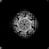
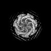
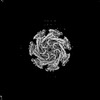
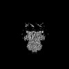
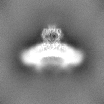
| Projections & slices | Image control
Images are generated by Spider. | ||||||||||||||||||||||||||||||||||||
| Voxel size | X=Y=Z: 0.94 Å | ||||||||||||||||||||||||||||||||||||
| Density |
| ||||||||||||||||||||||||||||||||||||
| Symmetry | Space group: 1 | ||||||||||||||||||||||||||||||||||||
| Details | EMDB XML:
|
 Z (Sec.)
 Y (Row.)
 X (Col.)


-Supplemental data
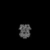
-Half map: Half A
| File | emd_26876_half_map_1.map | ||||||||||||
|---|---|---|---|---|---|---|---|---|---|---|---|---|---|
| Annotation | Half A | ||||||||||||
| Projections & Slices |
| ||||||||||||
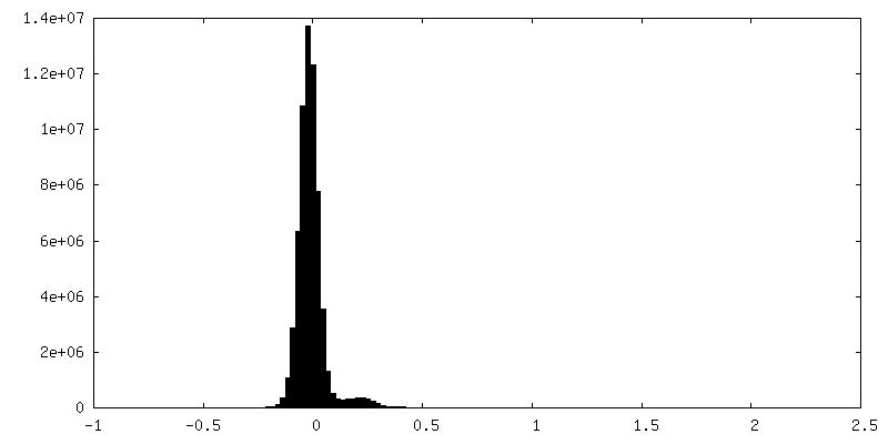
| Density Histograms |


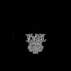
-Half map: Half B
| File | emd_26876_half_map_2.map | ||||||||||||
|---|---|---|---|---|---|---|---|---|---|---|---|---|---|
| Annotation | Half B | ||||||||||||
| Projections & Slices |
| ||||||||||||
| Density Histograms |


- Sample components
-Entire : EcMscK in an intermediate conformation
| Entire | Name: EcMscK in an intermediate conformation |
|---|---|
| Components |
|
-Supramolecule #1: EcMscK in an intermediate conformation
| Supramolecule | Name: EcMscK in an intermediate conformation / type: complex / Chimera: Yes / ID: 1 / Parent: 0 / Macromolecule list: all / Details: Homomeric Heptamer |
|---|---|
| Source (natural) | Organism: |
| Recombinant expression | Organism:  Komagataella pastoris (fungus) Komagataella pastoris (fungus) |
-Macromolecule #1: EcMscK
| Macromolecule | Name: EcMscK / type: protein_or_peptide / ID: 1 / Enantiomer: LEVO |
|---|---|
| Source (natural) | Organism: |
| Sequence | String: MTMFQYYKRS RHFVFSAFIA FVFVLLCQNT AFARASSNGD LPTKADLQAQ LDSLNKQKDL SAQDKLVQQ DLTDTLATLD KIDRIKEETV QLRQKVAEAP EKMRQATAAL TALSDVDNDE E TRKILSTL SLRQLETRVA QALDDLQNAQ NDLASYNSQL VSLQTQPERV ...String: MTMFQYYKRS RHFVFSAFIA FVFVLLCQNT AFARASSNGD LPTKADLQAQ LDSLNKQKDL SAQDKLVQQ DLTDTLATLD KIDRIKEETV QLRQKVAEAP EKMRQATAAL TALSDVDNDE E TRKILSTL SLRQLETRVA QALDDLQNAQ NDLASYNSQL VSLQTQPERV QNAMYNASQQ LQ QIRSRLD GTDVGETALR PSQKVLMQAQ QALLNAEIDQ QRKSLEGNTV LQDTLQKQRD YVT ANSARL EHQLQLLQEA VNSKRLTLTE KTAQEAVSPD EAARIQANPL VKQELEINQQ LSQR LITAT ENGNQLMQQN IKVKNWLERA LQSERNIKEQ IAVLKGSLLL SRILYQQQQT LPSAD ELEN MTNRIADLRL EQFEVNQQRD ALFQSDAFVN KLEEGHTNEV NSEVHDALLQ VVDMRR ELL DQLNKQLGNQ LMMAINLQIN QQQLMSVSKN LKSILTQQIF WVNSNRPMDW DWIKAFP QS LKDEFKSMKI TVNWQKAWPA VFIAFLAGLP LLLIAGLIHW RLGWLKAYQQ KLASAVGS L RNDSQLNTPK AILIDLIRAL PVCLIILAVG LILLTMQLNI SELLWSFSKK LAIFWLVFG LCWKVLEKNG VAVRHFGMPE QQTSHWRRQI VRISLALLPI HFWSVVAELS PLHLMDDVLG QAMIFFNLL LIAFLVWPMC RESWRDKESH TMRLVTITVL SIIPIALMVL TATGYFYTTL R LAGRWIET VYLVIIWNLL YQTVLRGLSV AARRIAWRRA LARRQNLVKE GAEGAEPPEE PT IALEQVN QQTLRITMLL MFALFGVMFW AIWSDLITVF SYLDSITLWH YNGTEAGAAV VKN VTMGSL LFAIIASMVA WALIRNLPGL LEVLVLSRLN MRQGASYAIT TILNYIIIAV GAMT VFGSL GVSWDKLQWL AAALSVGLSF GLQEIFGNFV SGLIILFERP VRIGDTVTIG SFSGT VSKI RIRATTITDF DRKEVIIPNK AFVTERLINW SLTDTTTRLV IRLGVAYGSD LEKVRK VLL KAATEHPRVM HEPMPEVFFT AFGASTLDHE LRLYVRELRD RSRTVDELNR TIDQLCR EN DINIAFNQLE VHLHNEKGDE VTEVKRDYKG DDPTPAVG |
-Experimental details
-Structure determination
| Method | cryo EM |
|---|---|
 Processing Processing | single particle reconstruction |
| Aggregation state | particle |
-Sample preparation
| Concentration | 7 mg/mL | |||||||||||||||
|---|---|---|---|---|---|---|---|---|---|---|---|---|---|---|---|---|
| Buffer | pH: 8 Component:
Details: Micrographs were pooled from protein purified in the presence of either 150 mM NaCl or 150 mM KCl. Buffers were prepared fresh, degassed, and filtered through a 0.45 um Durapore PVDF membrane. | |||||||||||||||
| Grid | Model: Quantifoil R2/2 / Material: COPPER / Mesh: 300 / Support film - Material: CARBON / Support film - topology: CONTINUOUS / Pretreatment - Type: GLOW DISCHARGE | |||||||||||||||
| Vitrification | Cryogen name: ETHANE / Chamber humidity: 100 % / Chamber temperature: 277.15 K / Instrument: FEI VITROBOT MARK IV | |||||||||||||||
| Details | The sample was homogeneous and monodisperse. |
- Electron microscopy
| Microscope | TFS GLACIOS |
|---|---|
| Image recording | Film or detector model: FEI FALCON IV (4k x 4k) / Average electron dose: 46.16 e/Å2 |
| Electron beam | Acceleration voltage: 200 kV / Electron source:  FIELD EMISSION GUN FIELD EMISSION GUN |
| Electron optics | Illumination mode: FLOOD BEAM / Imaging mode: BRIGHT FIELD / Nominal defocus max: 2.4 µm / Nominal defocus min: 0.6 µm / Nominal magnification: 150000 |
| Sample stage | Cooling holder cryogen: NITROGEN |
-Image processing
| Final reconstruction | Applied symmetry - Point group: C7 (7 fold cyclic) / Resolution.type: BY AUTHOR / Resolution: 3.85 Å / Resolution method: FSC 0.143 CUT-OFF / Number images used: 136195 |
|---|---|
| Initial angle assignment | Type: NOT APPLICABLE |
| Final angle assignment | Type: NOT APPLICABLE |
| FSC plot (resolution estimation) |  |
