Movie
Movie Controller

[English] 日本語
 Yorodumi
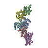
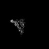
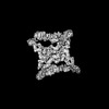
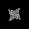
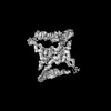
- PDB-7u05: Structure of the yeast TRAPPII-Rab11/Ypt32 complex in the closed/... -
+ Open data

- Basic information
| Entry | Database: PDB / ID: 7u05 | |||||||||
|---|---|---|---|---|---|---|---|---|---|---|
| Title | Structure of the yeast TRAPPII-Rab11/Ypt32 complex in the closed/closed state (composite structure) | |||||||||
 Components Components |
| |||||||||
 Keywords Keywords | PROTEIN TRANSPORT / complex / GTPase / Guanosine Exchange Factor / GEF | |||||||||
| Function / homology |  Function and homology information Function and homology informationAnchoring of the basal body to the plasma membrane / beta-glucan biosynthetic process / TRAPPI protein complex / RAB geranylgeranylation / RAB GEFs exchange GTP for GDP on RABs / TRAPPII protein complex / TRAPPIII protein complex / TRAPP complex / early endosome to Golgi transport / cytoplasm to vacuole targeting by the Cvt pathway ...Anchoring of the basal body to the plasma membrane / beta-glucan biosynthetic process / TRAPPI protein complex / RAB geranylgeranylation / RAB GEFs exchange GTP for GDP on RABs / TRAPPII protein complex / TRAPPIII protein complex / TRAPP complex / early endosome to Golgi transport / cytoplasm to vacuole targeting by the Cvt pathway / COPII-mediated vesicle transport / protein localization to phagophore assembly site / cis-Golgi network membrane / cellular bud neck / cis-Golgi network / intra-Golgi vesicle-mediated transport / phagophore assembly site / retrograde transport, endosome to Golgi / cellular response to nitrogen starvation / protein-containing complex localization / exocytosis / positive regulation of macroautophagy / chromosome organization / endoplasmic reticulum to Golgi vesicle-mediated transport / vesicle-mediated transport / Neutrophil degranulation / macroautophagy / trans-Golgi network / cell wall organization / recycling endosome / autophagy / protein transport / protein-containing complex assembly / early endosome / mitochondrial outer membrane / endosome / Golgi membrane / GTPase activity / GTP binding / endoplasmic reticulum / Golgi apparatus / nucleus / cytoplasm / cytosol Similarity search - Function | |||||||||
| Biological species |  | |||||||||
| Method | ELECTRON MICROSCOPY / single particle reconstruction / cryo EM / Resolution: 3.7 Å | |||||||||
 Authors Authors | Bagde, S.R. / Fromme, J.C. | |||||||||
| Funding support |  United States, 2items United States, 2items
| |||||||||
 Citation Citation | Journal: Sci Adv / Year: 2022 Title: Structure of a TRAPPII-Rab11 activation intermediate reveals GTPase substrate selection mechanisms. Authors: Saket R Bagde / J Christopher Fromme / Abstract: Rab1 and Rab11 are essential regulators of the eukaryotic secretory and endocytic recycling pathways. The transport protein particle (TRAPP) complexes activate these guanosine triphosphatases via ...Rab1 and Rab11 are essential regulators of the eukaryotic secretory and endocytic recycling pathways. The transport protein particle (TRAPP) complexes activate these guanosine triphosphatases via nucleotide exchange using a shared set of core subunits. The basal specificity of the TRAPP core is toward Rab1, yet the TRAPPII complex is specific for Rab11. A steric gating mechanism has been proposed to explain TRAPPII counterselection against Rab1. Here, we present cryo-electron microscopy structures of the 22-subunit TRAPPII complex from budding yeast, including a TRAPPII-Rab11 nucleotide exchange intermediate. The Trs130 subunit provides a "leg" that positions the active site distal to the membrane surface, and this leg is required for steric gating. The related TRAPPIII complex is unable to activate Rab11 because of a repulsive interaction, which TRAPPII surmounts using the Trs120 subunit as a "lid" to enclose the active site. TRAPPII also adopts an open conformation enabling Rab11 to access and exit from the active site chamber. | |||||||||
| History |
|
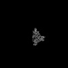
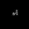
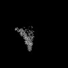
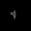
- Structure visualization
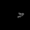
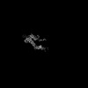
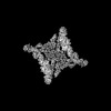
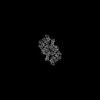
| Structure viewer | Molecule: MolmilJmol/JSmol |
|---|
- Downloads & links
-Download
| PDBx/mmCIF format | 7u05.cif.gz | 2.7 MB | Display | PDBx/mmCIF format |
|---|---|---|---|---|
| PDB format | pdb7u05.ent.gz | Display | PDB format | |
| PDBx/mmJSON format | 7u05.json.gz | Tree view | PDBx/mmJSON format | |
| Others |  Other downloads Other downloads |
-Validation report
| Arichive directory | https://data.pdbj.org/pub/pdb/validation_reports/u0/7u05ftp://data.pdbj.org/pub/pdb/validation_reports/u0/7u05 | HTTPS FTP |
|---|
-Related structure data
| Related structure data |  26254MC 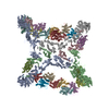 7u06C C: citing same article ( M: map data used to model this data |
|---|---|
| Similar structure data |


-Links
 PDBj
PDBj


- Assembly
| Deposited unit | 
|
|---|---|
| 1 |
|
-Components
-Trafficking protein particle complex II-specific subunit ... , 5 types, 10 molecules MmCcBbAaNn
| #1: Protein | Mass: 20613.320 Da / Num. of mol.: 2 / Fragment: N-terminal region / Source method: isolated from a natural source / Source: (natural) #2: Protein | Mass: 63434.473 Da / Num. of mol.: 2 / Source method: isolated from a natural source / Source: (natural) #11: Protein | Mass: 128424.250 Da / Num. of mol.: 2 / Source method: isolated from a natural source / Source: (natural) #12: Protein | Mass: 147823.281 Da / Num. of mol.: 2 / Source method: isolated from a natural source / Source: (natural) #13: Protein | Mass: 17889.996 Da / Num. of mol.: 2 / Fragment: N-terminal region / Source method: isolated from a natural source / Source: (natural) |
|---|
-Protein , 2 types, 4 molecules DdLl
| #3: Protein | Mass: 17371.008 Da / Num. of mol.: 2 / Source method: isolated from a natural source / Source: (natural) #10: Protein | Mass: 25309.145 Da / Num. of mol.: 2 Source method: isolated from a genetically manipulated source Source: (gene. exp.) Gene: YPT32, YPT11, YGL210W / Production host:  |
|---|
-Trafficking protein particle complex subunit ... , 6 types, 14 molecules EeFIfiGgHhJjKk
| #4: Protein | Mass: 30786.176 Da / Num. of mol.: 2 / Source method: isolated from a natural source / Source: (natural) #5: Protein | Mass: 22152.445 Da / Num. of mol.: 4 / Source method: isolated from a natural source / Source: (natural) #6: Protein | Mass: 18453.875 Da / Num. of mol.: 2 / Source method: isolated from a natural source / Source: (natural) #7: Protein | Mass: 24889.262 Da / Num. of mol.: 2 / Source method: isolated from a natural source / Source: (natural) #8: Protein | Mass: 31755.689 Da / Num. of mol.: 2 / Source method: isolated from a natural source / Source: (natural) #9: Protein | Mass: 19721.154 Da / Num. of mol.: 2 / Source method: isolated from a natural source / Source: (natural) |
|---|
-Non-polymers , 1 types, 4 molecules 
| #14: Chemical | ChemComp-PLM /  Mass: 256.424 Da / Num. of mol.: 4 / Source method: obtained synthetically / Formula: C16H32O2 / Feature type: SUBJECT OF INVESTIGATION Mass: 256.424 Da / Num. of mol.: 4 / Source method: obtained synthetically / Formula: C16H32O2 / Feature type: SUBJECT OF INVESTIGATION |
|---|
-Details
| Has ligand of interest | Y |
|---|---|
| Has protein modification | Y |
-Experimental details
-Experiment
| Experiment | Method: ELECTRON MICROSCOPY |
|---|---|
| EM experiment | Aggregation state: PARTICLE / 3D reconstruction method: single particle reconstruction |
- Sample preparation
| Component | Name: TRAPPII complex bound to Rab11/Ypt32 / Type: COMPLEX / Entity ID: #1-#13 / Source: MULTIPLE SOURCES | ||||||||||||||||||||||||
|---|---|---|---|---|---|---|---|---|---|---|---|---|---|---|---|---|---|---|---|---|---|---|---|---|---|
| Molecular weight | Value: 1.1 MDa / Experimental value: NO | ||||||||||||||||||||||||
| Source (natural) | Organism: | ||||||||||||||||||||||||
| Source (recombinant) | Organism: | ||||||||||||||||||||||||
| Buffer solution | pH: 8 | ||||||||||||||||||||||||
| Buffer component |
| ||||||||||||||||||||||||
| Specimen | Conc.: 5.6 mg/ml / Embedding applied: NO / Shadowing applied: NO / Staining applied: NO / Vitrification applied: YES | ||||||||||||||||||||||||
| Specimen support | Grid material: GOLD / Grid mesh size: 300 divisions/in. / Grid type: UltrAuFoil R1.2/1.3 | ||||||||||||||||||||||||
| Vitrification | Instrument: FEI VITROBOT MARK IV / Cryogen name: ETHANE / Humidity: 100 % / Chamber temperature: 277 K Details: The sample was incubated on the grid for 10 seconds followed by blotting for 5 seconds before plunging in liquid ethane. |
- Electron microscopy imaging
| Experimental equipment |  Model: Talos Arctica / Image courtesy: FEI Company |
|---|---|
| Microscopy | Model: FEI TALOS ARCTICA |
| Electron gun | Electron source:  FIELD EMISSION GUN / Accelerating voltage: 200 kV / Illumination mode: FLOOD BEAM FIELD EMISSION GUN / Accelerating voltage: 200 kV / Illumination mode: FLOOD BEAM |
| Electron lens | Mode: BRIGHT FIELD / Nominal magnification: 63000 X / Nominal defocus max: 2500 nm / Nominal defocus min: 800 nm / Cs: 2.7 mm / C2 aperture diameter: 50 µm / Alignment procedure: COMA FREE |
| Specimen holder | Cryogen: NITROGEN / Specimen holder model: FEI TITAN KRIOS AUTOGRID HOLDER |
| Image recording | Average exposure time: 3.5 sec. / Electron dose: 53 e/Å2 / Film or detector model: GATAN K3 BIOQUANTUM (6k x 4k) / Num. of grids imaged: 3 / Num. of real images: 4998 / Details: Images were collected as 50 frame movies. |
| EM imaging optics | Energyfilter name: GIF Bioquantum / Energyfilter slit width: 20 eV |
| Image scans | Width: 5760 / Height: 4092 |
- Processing
| Software |
| |||||||||||||||||||||||||||||||||||||||||||||
|---|---|---|---|---|---|---|---|---|---|---|---|---|---|---|---|---|---|---|---|---|---|---|---|---|---|---|---|---|---|---|---|---|---|---|---|---|---|---|---|---|---|---|---|---|---|---|
| EM software |
| |||||||||||||||||||||||||||||||||||||||||||||
| CTF correction | Type: PHASE FLIPPING AND AMPLITUDE CORRECTION | |||||||||||||||||||||||||||||||||||||||||||||
| Particle selection | Num. of particles selected: 979187 | |||||||||||||||||||||||||||||||||||||||||||||
| Symmetry | Point symmetry: C2 (2 fold cyclic) | |||||||||||||||||||||||||||||||||||||||||||||
| 3D reconstruction | Resolution: 3.7 Å / Resolution method: FSC 0.143 CUT-OFF / Num. of particles: 369488 Details: The composite map was generated by combining the consensus dimer map (EMD-26221) with focused refinement maps deposited in the entries EMD-26223, EMD-26224. EMD-26225, EMD-26226, EMD-26227, ...Details: The composite map was generated by combining the consensus dimer map (EMD-26221) with focused refinement maps deposited in the entries EMD-26223, EMD-26224. EMD-26225, EMD-26226, EMD-26227, EMD-26228, EMD-26229, EMD-26230, EMD-26231 and EMD-26232 using Combine Focused Maps in Phenix. Symmetry type: POINT | |||||||||||||||||||||||||||||||||||||||||||||
| Atomic model building | Protocol: OTHER / Space: REAL Details: Model was rebuilt in coot and refined using phenix.real_space_refine. | |||||||||||||||||||||||||||||||||||||||||||||
| Atomic model building |
| |||||||||||||||||||||||||||||||||||||||||||||
| Refinement | Cross valid method: NONE Stereochemistry target values: GeoStd + Monomer Library + CDL v1.2 | |||||||||||||||||||||||||||||||||||||||||||||
| Displacement parameters | Biso mean: 107.67 Å2 | |||||||||||||||||||||||||||||||||||||||||||||
| Refine LS restraints |
|