 Movie
Movie Controller

[English] 日本語
 Yorodumi
- PDB-7tgg: Cryo-EM structure of PilA-N and PilA-C from Geobacter sulfurreducens -
+ Open data

- Basic information
| Entry | Database: PDB / ID: 7tgg | |||||||||||||||||||||||||||
|---|---|---|---|---|---|---|---|---|---|---|---|---|---|---|---|---|---|---|---|---|---|---|---|---|---|---|---|---|
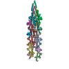
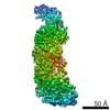
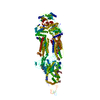
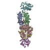
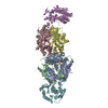
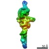
| Title | Cryo-EM structure of PilA-N and PilA-C from Geobacter sulfurreducens | |||||||||||||||||||||||||||
 Components Components |
| |||||||||||||||||||||||||||
 Keywords Keywords | PROTEIN FIBRIL / helical symmetry / filament / pili / type iv pili / pseudo pili | |||||||||||||||||||||||||||
| Function / homology |  Function and homology information Function and homology informationpilus assembly / protein secretion by the type II secretion system / type II protein secretion system complex / membrane Similarity search - Function | |||||||||||||||||||||||||||
| Biological species |  Geobacter sulfurreducens (bacteria) Geobacter sulfurreducens (bacteria) | |||||||||||||||||||||||||||
| Method | ELECTRON MICROSCOPY / helical reconstruction / cryo EM / Resolution: 4.1 Å | |||||||||||||||||||||||||||
 Authors Authors | Wang, F. / Mustafa, K. / Chan, C.H. / Joshi, K. / Bond, D.R. / Hochbaum, A.I. / Egelman, E.H. | |||||||||||||||||||||||||||
| Funding support |  United States, 2items United States, 2items
| |||||||||||||||||||||||||||
 Citation Citation | Journal: Nat Microbiol / Year: 2022 Title: Cryo-EM structure of an extracellular Geobacter OmcE cytochrome filament reveals tetrahaem packing. Authors: Fengbin Wang / Khawla Mustafa / Victor Suciu / Komal Joshi / Chi H Chan / Sol Choi / Zhangli Su / Dong Si / Allon I Hochbaum / Edward H Egelman / Daniel R Bond / Abstract: Electrically conductive appendages from the anaerobic bacterium Geobacter sulfurreducens were first observed two decades ago, with genetic and biochemical data suggesting that conductive fibres were ...Electrically conductive appendages from the anaerobic bacterium Geobacter sulfurreducens were first observed two decades ago, with genetic and biochemical data suggesting that conductive fibres were type IV pili. Recently, an extracellular conductive filament of G. sulfurreducens was found to contain polymerized c-type cytochrome OmcS subunits, not pilin subunits. Here we report that G. sulfurreducens also produces a second, thinner appendage comprised of cytochrome OmcE subunits and solve its structure using cryo-electron microscopy at ~4.3 Å resolution. Although OmcE and OmcS subunits have no overall sequence or structural similarities, upon polymerization both form filaments that share a conserved haem packing arrangement in which haems are coordinated by histidines in adjacent subunits. Unlike OmcS filaments, OmcE filaments are highly glycosylated. In extracellular fractions from G. sulfurreducens, we detected type IV pili comprising PilA-N and -C chains, along with abundant B-DNA. OmcE is the second cytochrome filament to be characterized using structural and biophysical methods. We propose that there is a broad class of conductive bacterial appendages with conserved haem packing (rather than sequence homology) that enable long-distance electron transport to chemicals or other microbial cells. | |||||||||||||||||||||||||||
| History |
|
- Structure visualization
| Movie |
Movie viewer |
|---|---|
| Structure viewer | Molecule: MolmilJmol/JSmol |
- Downloads & links
-Download
| PDBx/mmCIF format | 7tgg.cif.gz | 40.8 KB | Display | PDBx/mmCIF format |
|---|---|---|---|---|
| PDB format | pdb7tgg.ent.gz | 25.8 KB | Display | PDB format |
| PDBx/mmJSON format | 7tgg.json.gz | Tree view | PDBx/mmJSON format | |
| Others |  Other downloads Other downloads |
-Validation report
| Arichive directory | https://data.pdbj.org/pub/pdb/validation_reports/tg/7tggftp://data.pdbj.org/pub/pdb/validation_reports/tg/7tgg | HTTPS FTP |
|---|
-Related structure data
| Related structure data |  25881MC  7tfsC M: map data used to model this data C: citing same article ( |
|---|---|
| Similar structure data |


-Links
 PDBj
PDBj
- Assembly
| Deposited unit | 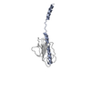
|
|---|---|
| 1 | x 15
|
-Components
| #1: Protein | Mass: 10006.582 Da / Num. of mol.: 1 / Source method: isolated from a natural source / Source: (natural) Geobacter sulfurreducens (bacteria) / Strain: ATCC 51573 / DSM 12127 / PCA / References: UniProt: Q74D23 |
|---|---|
| #2: Protein | Mass: 13087.543 Da / Num. of mol.: 1 / Source method: isolated from a natural source / Source: (natural) Geobacter sulfurreducens (bacteria) / Strain: ATCC 51573 / DSM 12127 / PCA / References: UniProt: Q74D22 |
| Has protein modification | N |
-Experimental details
-Experiment
| Experiment | Method: ELECTRON MICROSCOPY |
|---|---|
| EM experiment | Aggregation state: FILAMENT / 3D reconstruction method: helical reconstruction |
- Sample preparation
| Component | Name: Filament of PilA-N and PilA-C proteins / Type: COMPLEX / Entity ID: all / Source: NATURAL |
|---|---|
| Source (natural) | Organism: Geobacter sulfurreducens (bacteria) / Strain: ATCC 51573 / DSM 12127 / PCA |
| Buffer solution | pH: 6 |
| Specimen | Embedding applied: NO / Shadowing applied: NO / Staining applied: NO / Vitrification applied: YES |
| Vitrification | Cryogen name: ETHANE |
- Electron microscopy imaging
| Experimental equipment |  Model: Titan Krios / Image courtesy: FEI Company |
|---|---|
| Microscopy | Model: FEI TITAN KRIOS |
| Electron gun | Electron source:  FIELD EMISSION GUN / Accelerating voltage: 300 kV / Illumination mode: FLOOD BEAM FIELD EMISSION GUN / Accelerating voltage: 300 kV / Illumination mode: FLOOD BEAM |
| Electron lens | Mode: BRIGHT FIELD / Nominal defocus max: 3000 nm / Nominal defocus min: 1000 nm |
| Image recording | Electron dose: 50 e/Å2 / Film or detector model: GATAN K3 (6k x 4k) |
- Processing
| Software | Name: PHENIX / Version: 1.19.2_4158: / Classification: refinement | ||||||||||||||||||||||||
|---|---|---|---|---|---|---|---|---|---|---|---|---|---|---|---|---|---|---|---|---|---|---|---|---|---|
| EM software | Name: PHENIX / Category: model refinement | ||||||||||||||||||||||||
| CTF correction | Type: PHASE FLIPPING AND AMPLITUDE CORRECTION | ||||||||||||||||||||||||
| Helical symmerty | Angular rotation/subunit: 89.1 ° / Axial rise/subunit: 10.4 Å / Axial symmetry: C1 | ||||||||||||||||||||||||
| 3D reconstruction | Resolution: 4.1 Å / Resolution method: FSC 0.143 CUT-OFF / Num. of particles: 112011 / Symmetry type: HELICAL | ||||||||||||||||||||||||
| Refine LS restraints |
|
