 Movie
Movie Controller

[English] 日本語
 Yorodumi
- PDB-6gv9: Structure of the type IV pilus from enterohemorrhagic Escherichia... -
+ Open data

- Basic information
| Entry | Database: PDB / ID: 6gv9 | ||||||
|---|---|---|---|---|---|---|---|
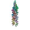
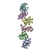
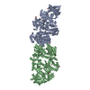
| Title | Structure of the type IV pilus from enterohemorrhagic Escherichia coli (EHEC) | ||||||
 Components Components | Prepilin peptidase-dependent protein D | ||||||
 Keywords Keywords | PROTEIN FIBRIL / TYPE IV PILI EHEC T4P CELL ADHESION hemorrhagic coli pilus (HCP) | ||||||
| Function / homology | type IV pilus / type IV pilus-dependent motility / Prokaryotic N-terminal methylation site. / Prokaryotic N-terminal methylation motif / Prokaryotic N-terminal methylation site / Pilin-like / membrane / Prepilin peptidase-dependent pilin Function and homology information Function and homology information | ||||||
| Biological species |  | ||||||
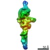
| Method | ELECTRON MICROSCOPY / helical reconstruction / cryo EM / Resolution: 8 Å | ||||||
 Authors Authors | Bardiaux, B. / Amorim, G.C. / Luna-Rico, A. / Zheng, W. / Guilvout, I. / Jollivet, C. / Nilges, M. / Egelman, E. / Francetic, O. / Izadi-Pruneyre, N. | ||||||
| Funding support |  France, 1items France, 1items
| ||||||
 Citation Citation | Journal: Structure / Year: 2019 Title: Structure and Assembly of the Enterohemorrhagic Escherichia coli Type 4 Pilus. Authors: Benjamin Bardiaux / Gisele Cardoso de Amorim / Areli Luna Rico / Weili Zheng / Ingrid Guilvout / Camille Jollivet / Michael Nilges / Edward H Egelman / Nadia Izadi-Pruneyre / Olivera Francetic /  Abstract: Bacterial type 4a pili are dynamic surface filaments that promote bacterial adherence, motility, and macromolecular transport. Their genes are highly conserved among enterobacteria and their ...Bacterial type 4a pili are dynamic surface filaments that promote bacterial adherence, motility, and macromolecular transport. Their genes are highly conserved among enterobacteria and their expression in enterohemorrhagic Escherichia coli (EHEC) promotes adhesion to intestinal epithelia and pro-inflammatory signaling. To define the molecular basis of EHEC pilus assembly, we determined the structure of the periplasmic domain of its major subunit PpdD (PpdDp), a prototype of an enterobacterial pilin subfamily containing two disulfide bonds. The structure of PpdDp, determined by NMR, was then docked into the density envelope of purified EHEC pili obtained by cryoelectron microscopy (cryo-EM). Cryo-EM reconstruction of EHEC pili at ∼8 Å resolution revealed extremely high pilus flexibility correlating with a large extended region of the pilin stem. Systematic mutagenesis combined with functional and interaction analyses identified charged residues essential for pilus assembly. Structural information on exposed regions and interfaces between EHEC pilins is relevant for vaccine and drug discovery. | ||||||
| History |
|
- Structure visualization
| Movie |
Movie viewer |
|---|---|
| Structure viewer | Molecule: MolmilJmol/JSmol |
- Downloads & links
-Download
| PDBx/mmCIF format | 6gv9.cif.gz | 582.2 KB | Display | PDBx/mmCIF format |
|---|---|---|---|---|
| PDB format | pdb6gv9.ent.gz | 480.8 KB | Display | PDB format |
| PDBx/mmJSON format | 6gv9.json.gz | Tree view | PDBx/mmJSON format | |
| Others |  Other downloads Other downloads |
-Validation report
| Arichive directory | https://data.pdbj.org/pub/pdb/validation_reports/gv/6gv9ftp://data.pdbj.org/pub/pdb/validation_reports/gv/6gv9 | HTTPS FTP |
|---|
-Related structure data
| Related structure data | 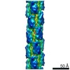 0070MC 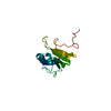 6gmsC C: citing same article ( M: map data used to model this data |
|---|---|
| Similar structure data |


-Links
 PDBj
PDBj
- Assembly
| Deposited unit | 
|
|---|---|
| 1 |
|
-Components
| #1: Protein | Mass: 14883.821 Da / Num. of mol.: 14 Source method: isolated from a genetically manipulated source Source: (gene. exp.) Has protein modification | Y | |
|---|
-Experimental details
-Experiment
| Experiment | Method: ELECTRON MICROSCOPY |
|---|---|
| EM experiment | Aggregation state: FILAMENT / 3D reconstruction method: helical reconstruction |
- Sample preparation
| Component | Name: EHEC type IV pili / Type: COMPLEX / Entity ID: all / Source: RECOMBINANT |
|---|---|
| Molecular weight | Experimental value: NO |
| Source (natural) | Organism: |
| Source (recombinant) | Organism: |
| Buffer solution | pH: 7.4 |
| Specimen | Embedding applied: NO / Shadowing applied: NO / Staining applied: NO / Vitrification applied: YES |
| Specimen support | Grid material: COPPER / Grid mesh size: 300 divisions/in. |
| Vitrification | Instrument: FEI VITROBOT MARK IV / Cryogen name: ETHANE / Humidity: 95 % / Chamber temperature: 297 K |
- Electron microscopy imaging
| Experimental equipment |  Model: Titan Krios / Image courtesy: FEI Company |
|---|---|
| Microscopy | Model: FEI TITAN KRIOS |
| Electron gun | Electron source:  FIELD EMISSION GUN / Accelerating voltage: 300 kV / Illumination mode: FLOOD BEAM FIELD EMISSION GUN / Accelerating voltage: 300 kV / Illumination mode: FLOOD BEAM |
| Electron lens | Mode: BRIGHT FIELD |
| Image recording | Average exposure time: 2 sec. / Electron dose: 1.4 e/Å2 / Detector mode: INTEGRATING / Film or detector model: FEI FALCON III (4k x 4k) / Num. of grids imaged: 1 |
- Processing
| EM software |
| ||||||||||||||||||||
|---|---|---|---|---|---|---|---|---|---|---|---|---|---|---|---|---|---|---|---|---|---|
| CTF correction | Type: PHASE FLIPPING AND AMPLITUDE CORRECTION | ||||||||||||||||||||
| Helical symmerty | Angular rotation/subunit: 96 ° / Axial rise/subunit: 11.2 Å / Axial symmetry: C1 | ||||||||||||||||||||
| 3D reconstruction | Resolution: 8 Å / Resolution method: FSC 0.143 CUT-OFF / Num. of particles: 25669 / Symmetry type: HELICAL | ||||||||||||||||||||
| Atomic model building | Protocol: OTHER / Space: REAL Details: Soluble domain structure and homology model of TM segment fitted as rigid-bodies. Linker region built ab initio. Real-space refinement after symmetrization. |
