 Movie
Movie Controller

[English] 日本語
 Yorodumi
- PDB-7sss: Structure of the NADH-bound human COQ7:COQ9 complex by single-par... -
+ Open data

- Basic information
| Entry | Database: PDB / ID: 7sss | |||||||||||||||||||||||||||||||||||||||||||||||||||||||||||||||||||||||||||||||||||||||||||||
|---|---|---|---|---|---|---|---|---|---|---|---|---|---|---|---|---|---|---|---|---|---|---|---|---|---|---|---|---|---|---|---|---|---|---|---|---|---|---|---|---|---|---|---|---|---|---|---|---|---|---|---|---|---|---|---|---|---|---|---|---|---|---|---|---|---|---|---|---|---|---|---|---|---|---|---|---|---|---|---|---|---|---|---|---|---|---|---|---|---|---|---|---|---|---|
| Title | Structure of the NADH-bound human COQ7:COQ9 complex by single-particle electron cryo-microscopy | |||||||||||||||||||||||||||||||||||||||||||||||||||||||||||||||||||||||||||||||||||||||||||||
 Components Components |
| |||||||||||||||||||||||||||||||||||||||||||||||||||||||||||||||||||||||||||||||||||||||||||||
 Keywords Keywords | MEMBRANE PROTEIN / hydroxylase / complex / lipid / enzyme / NADH / coenzymeQ / isoprene / OPP | |||||||||||||||||||||||||||||||||||||||||||||||||||||||||||||||||||||||||||||||||||||||||||||
| Function / homology |  Function and homology information Function and homology information3-demethoxyubiquinone 3-hydroxylase (NADH) / 3-demethoxyubiquinone 3-hydroxylase (NADH) activity / Ubiquinol biosynthesis / ubiquinone biosynthesis complex / extrinsic component of mitochondrial inner membrane / isoprenoid binding / ubiquinone biosynthetic process / regulation of reactive oxygen species metabolic process / mitochondrial electron transport, NADH to ubiquinone / determination of adult lifespan ...3-demethoxyubiquinone 3-hydroxylase (NADH) / 3-demethoxyubiquinone 3-hydroxylase (NADH) activity / Ubiquinol biosynthesis / ubiquinone biosynthesis complex / extrinsic component of mitochondrial inner membrane / isoprenoid binding / ubiquinone biosynthetic process / regulation of reactive oxygen species metabolic process / mitochondrial electron transport, NADH to ubiquinone / determination of adult lifespan / enzyme activator activity / chromosome / regulation of gene expression / mitochondrial inner membrane / chromatin binding / lipid binding / negative regulation of transcription by RNA polymerase II / protein homodimerization activity / positive regulation of transcription by RNA polymerase II / mitochondrion / metal ion binding / nucleus Similarity search - Function | |||||||||||||||||||||||||||||||||||||||||||||||||||||||||||||||||||||||||||||||||||||||||||||
| Biological species |  Homo sapiens (human) Homo sapiens (human) | |||||||||||||||||||||||||||||||||||||||||||||||||||||||||||||||||||||||||||||||||||||||||||||
| Method | ELECTRON MICROSCOPY / single particle reconstruction / cryo EM / Resolution: 2.4 Å | |||||||||||||||||||||||||||||||||||||||||||||||||||||||||||||||||||||||||||||||||||||||||||||
 Authors Authors | Aydin, H. / Frost, A. | |||||||||||||||||||||||||||||||||||||||||||||||||||||||||||||||||||||||||||||||||||||||||||||
| Funding support |  United States, 1items United States, 1items
| |||||||||||||||||||||||||||||||||||||||||||||||||||||||||||||||||||||||||||||||||||||||||||||
 Citation Citation | Journal: Mol Cell / Year: 2022 Title: Structure and functionality of a multimeric human COQ7:COQ9 complex. Authors: Mateusz Manicki / Halil Aydin / Luciano A Abriata / Katherine A Overmyer / Rachel M Guerra / Joshua J Coon / Matteo Dal Peraro / Adam Frost / David J Pagliarini /  Abstract: Coenzyme Q (CoQ) is a redox-active lipid essential for core metabolic pathways and antioxidant defense. CoQ is synthesized upon the mitochondrial inner membrane by an ill-defined "complex Q" ...Coenzyme Q (CoQ) is a redox-active lipid essential for core metabolic pathways and antioxidant defense. CoQ is synthesized upon the mitochondrial inner membrane by an ill-defined "complex Q" metabolon. Here, we present structure-function analyses of a lipid-, substrate-, and NADH-bound complex comprising two complex Q subunits: the hydroxylase COQ7 and the lipid-binding protein COQ9. We reveal that COQ7 adopts a ferritin-like fold with a hydrophobic channel whose substrate-binding capacity is enhanced by COQ9. Using molecular dynamics, we further show that two COQ7:COQ9 heterodimers form a curved tetramer that deforms the membrane, potentially opening a pathway for the CoQ intermediates to translocate from the bilayer to the proteins' lipid-binding sites. Two such tetramers assemble into a soluble octamer with a pseudo-bilayer of lipids captured within. Together, these observations indicate that COQ7 and COQ9 cooperate to access hydrophobic precursors within the membrane and coordinate subsequent synthesis steps toward producing CoQ. | |||||||||||||||||||||||||||||||||||||||||||||||||||||||||||||||||||||||||||||||||||||||||||||
| History |
|
- Structure visualization
| Structure viewer | Molecule: MolmilJmol/JSmol |
|---|
- Downloads & links
-Download
| PDBx/mmCIF format | 7sss.cif.gz | 332.6 KB | Display | PDBx/mmCIF format |
|---|---|---|---|---|
| PDB format | pdb7sss.ent.gz | 254.3 KB | Display | PDB format |
| PDBx/mmJSON format | 7sss.json.gz | Tree view | PDBx/mmJSON format | |
| Others |  Other downloads Other downloads |
-Validation report
| Arichive directory | https://data.pdbj.org/pub/pdb/validation_reports/ss/7sssftp://data.pdbj.org/pub/pdb/validation_reports/ss/7sss | HTTPS FTP |
|---|
-Related structure data
| Related structure data |  25413MC  7sspC M: map data used to model this data C: citing same article ( |
|---|---|
| Similar structure data |

-Links
 PDBj
PDBj- Assembly
| Deposited unit | 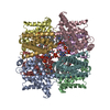
|
|---|---|
| 1 |
|
-Components
-Protein , 2 types, 8 molecules ABCDEFGH
| #1: Protein | Mass: 35549.945 Da / Num. of mol.: 4 Source method: isolated from a genetically manipulated source Source: (gene. exp.) Homo sapiens (human) / Gene: COQ9, C16orf49, HSPC326, PSEC0129 / Production host:  #2: Protein | Mass: 24313.064 Da / Num. of mol.: 4 Source method: isolated from a genetically manipulated source Source: (gene. exp.) Homo sapiens (human) / Gene: COQ7 / Production host: |
|---|
-Non-polymers , 4 types, 208 molecules 


| #3: Chemical | ChemComp-PEV / (  Mass: 720.012 Da / Num. of mol.: 32 / Source method: obtained synthetically / Formula: C39H78NO8P / Feature type: SUBJECT OF INVESTIGATION / Comment: POPE, phospholipid*YM Mass: 720.012 Da / Num. of mol.: 32 / Source method: obtained synthetically / Formula: C39H78NO8P / Feature type: SUBJECT OF INVESTIGATION / Comment: POPE, phospholipid*YM#4: Chemical | ChemComp-8PP /  Mass: 639.047 Da / Num. of mol.: 4 / Source method: obtained synthetically / Formula: C46H70O / Feature type: SUBJECT OF INVESTIGATION Mass: 639.047 Da / Num. of mol.: 4 / Source method: obtained synthetically / Formula: C46H70O / Feature type: SUBJECT OF INVESTIGATION#5: Chemical |  Mass: 663.425 Da / Num. of mol.: 2 / Source method: obtained synthetically / Formula: C21H27N7O14P2 / Comment: NAD*YM Mass: 663.425 Da / Num. of mol.: 2 / Source method: obtained synthetically / Formula: C21H27N7O14P2 / Comment: NAD*YM#6: Water | ChemComp-HOH / | Mass: 18.015 Da / Num. of mol.: 170 / Source method: isolated from a natural source / Formula: H2O |
|---|
-Details
| Has ligand of interest | Y |
|---|---|
| Has protein modification | N |
-Experimental details
-Experiment
| Experiment | Method: ELECTRON MICROSCOPY |
|---|---|
| EM experiment | Aggregation state: PARTICLE / 3D reconstruction method: single particle reconstruction |
- Sample preparation
| Component | Name: Human COQ7:COQ9 Complex / Type: COMPLEX / Entity ID: #1-#2 / Source: RECOMBINANT |
|---|---|
| Source (natural) | Organism: Homo sapiens (human) |
| Source (recombinant) | Organism: |
| Buffer solution | pH: 7 |
| Specimen | Embedding applied: NO / Shadowing applied: NO / Staining applied: NO / Vitrification applied: YES |
| Vitrification | Cryogen name: ETHANE |
- Electron microscopy imaging
| Experimental equipment |  Model: Titan Krios / Image courtesy: FEI Company |
|---|---|
| Microscopy | Model: FEI TITAN KRIOS |
| Electron gun | Electron source:  FIELD EMISSION GUN / Accelerating voltage: 300 kV / Illumination mode: FLOOD BEAM FIELD EMISSION GUN / Accelerating voltage: 300 kV / Illumination mode: FLOOD BEAM |
| Electron lens | Mode: BRIGHT FIELD / Nominal defocus max: 1200 nm / Nominal defocus min: 300 nm |
| Image recording | Electron dose: 65 e/Å2 / Film or detector model: GATAN K3 (6k x 4k) |
- Processing
| Software | Name: PHENIX / Version: 1.19.1_4122: / Classification: refinement | ||||||||||||||||||||||||
|---|---|---|---|---|---|---|---|---|---|---|---|---|---|---|---|---|---|---|---|---|---|---|---|---|---|
| EM software | Name: PHENIX / Category: model refinement | ||||||||||||||||||||||||
| CTF correction | Type: PHASE FLIPPING AND AMPLITUDE CORRECTION | ||||||||||||||||||||||||
| 3D reconstruction | Resolution: 2.4 Å / Resolution method: FSC 0.143 CUT-OFF / Num. of particles: 372917 / Symmetry type: POINT | ||||||||||||||||||||||||
| Refine LS restraints |
|
