 Movie
Movie Controller

[English] 日本語
 Yorodumi
- PDB-7rye: Cryo-EM structure of the needle filament-tip complex of the Salmo... -
+ Open data

- Basic information
| Entry | Database: PDB / ID: 7rye | |||||||||||||||||||||
|---|---|---|---|---|---|---|---|---|---|---|---|---|---|---|---|---|---|---|---|---|---|---|
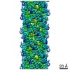
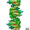
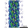
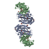
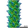
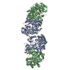
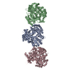
| Title | Cryo-EM structure of the needle filament-tip complex of the Salmonella type III secretion injectisome | |||||||||||||||||||||
 Components Components |
| |||||||||||||||||||||
 Keywords Keywords | CELL INVASION / protein secretion / bacterial pathogenesis / organelle assembly | |||||||||||||||||||||
| Function / homology |  Function and homology information Function and homology informationtype III protein secretion system complex / protein secretion by the type III secretion system / cell surface / extracellular region / identical protein binding Similarity search - Function | |||||||||||||||||||||
| Biological species |  Salmonella enterica subsp. enterica serovar Typhimurium (bacteria) Salmonella enterica subsp. enterica serovar Typhimurium (bacteria) | |||||||||||||||||||||
| Method | ELECTRON MICROSCOPY / single particle reconstruction / cryo EM / Resolution: 3.9 Å | |||||||||||||||||||||
 Authors Authors | Guo, E.Z. / Galan, J.E. | |||||||||||||||||||||
| Funding support |  United States, 1items United States, 1items
| |||||||||||||||||||||
 Citation Citation | Journal: Proc Natl Acad Sci U S A / Year: 2021 Title: Cryo-EM structure of the needle filament tip complex of the type III secretion injectisome. Authors: Emily Z Guo / Jorge E Galán / Abstract: Type III secretion systems are multiprotein molecular machines required for the virulence of several important bacterial pathogens. The central element of these machines is the injectisome, a ∼5-Md ...Type III secretion systems are multiprotein molecular machines required for the virulence of several important bacterial pathogens. The central element of these machines is the injectisome, a ∼5-Md multiprotein structure that mediates the delivery of bacterially encoded proteins into eukaryotic target cells. The injectisome is composed of a cytoplasmic sorting platform, and a membrane-embedded needle complex, which is made up of a multiring base and a needle-like filament that extends several nanometers from the bacterial surface. The needle filament is capped at its distal end by another substructure known as the tip complex, which is crucial for the translocation of effector proteins through the eukaryotic cell plasma membrane. Here we report the cryo-EM structure of the Typhimurium needle tip complex docked onto the needle filament tip. Combined with a detailed analysis of structurally guided mutants, this study provides major insight into the assembly and function of this essential component of the type III secretion protein injection machine. | |||||||||||||||||||||
| History |
|
- Structure visualization
| Movie |
Movie viewer |
|---|---|
| Structure viewer | Molecule: MolmilJmol/JSmol |
- Downloads & links
-Download
| PDBx/mmCIF format | 7rye.cif.gz | 429.8 KB | Display | PDBx/mmCIF format |
|---|---|---|---|---|
| PDB format | pdb7rye.ent.gz | 356.6 KB | Display | PDB format |
| PDBx/mmJSON format | 7rye.json.gz | Tree view | PDBx/mmJSON format | |
| Others |  Other downloads Other downloads |
-Validation report
| Arichive directory | https://data.pdbj.org/pub/pdb/validation_reports/ry/7ryeftp://data.pdbj.org/pub/pdb/validation_reports/ry/7rye | HTTPS FTP |
|---|
-Related structure data
| Related structure data |  24735MC M: map data used to model this data C: citing same article ( |
|---|---|
| Similar structure data |


-Links
 PDBj
PDBj- Assembly
| Deposited unit | 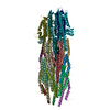
|
|---|---|
| 1 |
|
-Components
| #1: Protein | Mass: 8864.868 Da / Num. of mol.: 19 / Source method: isolated from a natural source Source: (natural) Salmonella enterica subsp. enterica serovar Typhimurium (bacteria)References: UniProt: P41784 #2: Protein | Mass: 37141.148 Da / Num. of mol.: 5 / Source method: isolated from a natural source Source: (natural) Salmonella enterica subsp. enterica serovar Typhimurium (bacteria)References: UniProt: A0A0C5PQX9 Has protein modification | N | |
|---|
-Experimental details
-Experiment
| Experiment | Method: ELECTRON MICROSCOPY |
|---|---|
| EM experiment | Aggregation state: PARTICLE / 3D reconstruction method: single particle reconstruction |
- Sample preparation
| Component | Name: The needle complex with tip / Type: COMPLEX / Entity ID: all / Source: NATURAL | |||||||||||||||
|---|---|---|---|---|---|---|---|---|---|---|---|---|---|---|---|---|
| Source (natural) | Organism: Salmonella enterica subsp. enterica serovar Typhimurium (bacteria) Strain: SL1344 | |||||||||||||||
| Buffer solution | pH: 7.5 | |||||||||||||||
| Buffer component |
| |||||||||||||||
| Specimen | Embedding applied: NO / Shadowing applied: NO / Staining applied: NO / Vitrification applied: YES | |||||||||||||||
| Specimen support | Details: 25 mAmp / Grid material: COPPER / Grid mesh size: 400 divisions/in. / Grid type: Quantifoil R2/2 | |||||||||||||||
| Vitrification | Instrument: FEI VITROBOT MARK IV / Cryogen name: ETHANE / Humidity: 100 % / Chamber temperature: 283 K |
- Electron microscopy imaging
| Experimental equipment |  Model: Titan Krios / Image courtesy: FEI Company |
|---|---|
| Microscopy | Model: FEI TITAN KRIOS |
| Electron gun | Electron source:  FIELD EMISSION GUN / Accelerating voltage: 300 kV / Illumination mode: FLOOD BEAM FIELD EMISSION GUN / Accelerating voltage: 300 kV / Illumination mode: FLOOD BEAM |
| Electron lens | Mode: BRIGHT FIELD / Cs: 2.7 mm / Alignment procedure: COMA FREE |
| Specimen holder | Cryogen: NITROGEN |
| Image recording | Electron dose: 50 e/Å2 / Detector mode: SUPER-RESOLUTION / Film or detector model: GATAN K2 SUMMIT (4k x 4k) |
- Processing
| Software | Name: PHENIX / Version: 1.19.2_4158: / Classification: refinement | ||||||||||||||||||||||||
|---|---|---|---|---|---|---|---|---|---|---|---|---|---|---|---|---|---|---|---|---|---|---|---|---|---|
| EM software |
| ||||||||||||||||||||||||
| CTF correction | Type: PHASE FLIPPING AND AMPLITUDE CORRECTION | ||||||||||||||||||||||||
| Symmetry | Point symmetry: C1 (asymmetric) | ||||||||||||||||||||||||
| 3D reconstruction | Resolution: 3.9 Å / Resolution method: FSC 0.143 CUT-OFF / Num. of particles: 27737 / Algorithm: BACK PROJECTION / Symmetry type: POINT | ||||||||||||||||||||||||
| Atomic model building | B value: 85 / Protocol: FLEXIBLE FIT | ||||||||||||||||||||||||
| Refine LS restraints |
|
