 Movie
Movie Controller

[English] 日本語
 Yorodumi
- PDB-7rro: Structure of the 48-nm repeat doublet microtubule from bovine tra... -
+ Open data

- Basic information
| Entry | Database: PDB / ID: 7rro | ||||||
|---|---|---|---|---|---|---|---|
| Title | Structure of the 48-nm repeat doublet microtubule from bovine tracheal cilia | ||||||
 Components Components |
| ||||||
 Keywords Keywords | STRUCTURAL PROTEIN / cilia / microtubule / dynein / motility | ||||||
| Function / homology |  Function and homology information Function and homology informationouter dynein arm docking complex / protein localization to motile cilium / regulation of cilium movement / mucociliary clearance / outer acrosomal membrane / epithelial cilium movement involved in determination of left/right asymmetry / regulation of brood size / establishment of left/right asymmetry / 9+0 motile cilium / sperm flagellum assembly ...outer dynein arm docking complex / protein localization to motile cilium / regulation of cilium movement / mucociliary clearance / outer acrosomal membrane / epithelial cilium movement involved in determination of left/right asymmetry / regulation of brood size / establishment of left/right asymmetry / 9+0 motile cilium / sperm flagellum assembly / manchette assembly / axonemal B tubule inner sheath / axonemal A tubule inner sheath / regulation of flagellated sperm motility / protein polyglutamylation / regulation of calcineurin-NFAT signaling cascade / Microtubule-dependent trafficking of connexons from Golgi to the plasma membrane / Cilium Assembly / Intraflagellar transport / : / Sealing of the nuclear envelope (NE) by ESCRT-III / Kinesins / positive regulation of feeding behavior / Resolution of Sister Chromatid Cohesion / Mitotic Prometaphase / EML4 and NUDC in mitotic spindle formation / outer dynein arm assembly / COPI-dependent Golgi-to-ER retrograde traffic / COPI-independent Golgi-to-ER retrograde traffic / COPI-mediated anterograde transport / RHO GTPases activate IQGAPs / inner dynein arm assembly / cilium-dependent cell motility / RHO GTPases Activate Formins / cerebrospinal fluid circulation / regulation of cilium beat frequency involved in ciliary motility / 9+2 motile cilium / MHC class II antigen presentation / cilium movement involved in cell motility / HSP90 chaperone cycle for steroid hormone receptors (SHR) in the presence of ligand / regulation of store-operated calcium entry / acrosomal membrane / regulation of cilium assembly / axoneme assembly / cilium movement / Aggrephagy / The role of GTSE1 in G2/M progression after G2 checkpoint / calcium ion sensor activity / Separation of Sister Chromatids / Loss of Nlp from mitotic centrosomes / Recruitment of mitotic centrosome proteins and complexes / Loss of proteins required for interphase microtubule organization from the centrosome / Anchoring of the basal body to the plasma membrane / AURKA Activation by TPX2 / axonemal microtubule / Recruitment of NuMA to mitotic centrosomes / cilium organization / gamma-tubulin ring complex / ciliary rootlet / Regulation of PLK1 Activity at G2/M Transition / manchette / Hedgehog 'off' state / 3'-5'-DNA exonuclease activity / flagellated sperm motility / UTP biosynthetic process / CTP biosynthetic process / motile cilium / determination of left/right symmetry / DNA catabolic process / Neutrophil degranulation / nucleoside diphosphate kinase activity / positive regulation of cell motility / GTP biosynthetic process / Hydrolases; Acting on ester bonds; Exodeoxyribonucleases producing 5'-phosphomonoesters / AMP binding / regulation of neuron projection development / cerebral cortex cell migration / ciliary transition zone / mitotic cytokinesis / microtubule organizing center / cellular response to UV-C / cilium assembly / regulation of cell division / spermatid development / regulation of signal transduction / single fertilization / axoneme / glial cell projection / sperm flagellum / alpha-tubulin binding / beta-tubulin binding / intercellular bridge / lung development / Hsp70 protein binding / ciliary tip / acrosomal vesicle / mitotic spindle organization / meiotic cell cycle / centriole / Hsp90 protein binding Similarity search - Function | ||||||
| Biological species |  | ||||||
| Method | ELECTRON MICROSCOPY / single particle reconstruction / cryo EM / Resolution: 3.4 Å | ||||||
 Authors Authors | Gui, M. / Anderson, J.R. / Botsch, J.J. / Meleppattu, S. / Singh, S.K. / Zhang, Q. / Brown, A. | ||||||
| Funding support |  United States, 1items United States, 1items
| ||||||
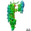
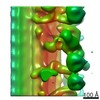
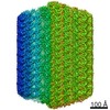
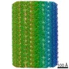
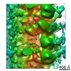
 Citation Citation | Journal: Cell / Year: 2021 Title: De novo identification of mammalian ciliary motility proteins using cryo-EM. Authors: Miao Gui / Hannah Farley / Priyanka Anujan / Jacob R Anderson / Dale W Maxwell / Jonathan B Whitchurch / J Josephine Botsch / Tao Qiu / Shimi Meleppattu / Sandeep K Singh / Qi Zhang / James ...Authors: Miao Gui / Hannah Farley / Priyanka Anujan / Jacob R Anderson / Dale W Maxwell / Jonathan B Whitchurch / J Josephine Botsch / Tao Qiu / Shimi Meleppattu / Sandeep K Singh / Qi Zhang / James Thompson / Jane S Lucas / Colin D Bingle / Dominic P Norris / Sudipto Roy / Alan Brown /   Abstract: Dynein-decorated doublet microtubules (DMTs) are critical components of the oscillatory molecular machine of cilia, the axoneme, and have luminal surfaces patterned periodically by microtubule inner ...Dynein-decorated doublet microtubules (DMTs) are critical components of the oscillatory molecular machine of cilia, the axoneme, and have luminal surfaces patterned periodically by microtubule inner proteins (MIPs). Here we present an atomic model of the 48-nm repeat of a mammalian DMT, derived from a cryoelectron microscopy (cryo-EM) map of the complex isolated from bovine respiratory cilia. The structure uncovers principles of doublet microtubule organization and features specific to vertebrate cilia, including previously unknown MIPs, a luminal bundle of tektin filaments, and a pentameric dynein-docking complex. We identify a mechanism for bridging 48- to 24-nm periodicity across the microtubule wall and show that loss of the proteins involved causes defective ciliary motility and laterality abnormalities in zebrafish and mice. Our structure identifies candidate genes for diagnosis of ciliopathies and provides a framework to understand their functions in driving ciliary motility. | ||||||
| History |
|
- Structure visualization
| Movie |
Movie viewer |
|---|---|
| Structure viewer | Molecule: MolmilJmol/JSmol |
- Downloads & links
-Download
| PDBx/mmCIF format | 7rro.cif.gz | 26.7 MB | Display | PDBx/mmCIF format |
|---|---|---|---|---|
| PDB format | pdb7rro.ent.gz | Display | PDB format | |
| PDBx/mmJSON format | 7rro.json.gz | Tree view | PDBx/mmJSON format | |
| Others |  Other downloads Other downloads |
-Validation report
| Arichive directory | https://data.pdbj.org/pub/pdb/validation_reports/rr/7rroftp://data.pdbj.org/pub/pdb/validation_reports/rr/7rro | HTTPS FTP |
|---|
-Related structure data
| Related structure data |  24664MC C: citing same article ( M: map data used to model this data |
|---|---|
| Similar structure data |
-Links
 PDBj
PDBj


- Assembly
| Deposited unit | 
|
|---|---|
| 1 |
|
-Components
+Protein , 27 types, 407 molecules 073456ABA0A1A2A3A4AAACAEAGAIAKAMBABCBEBGBIBKBMCACCCE...
-EF-hand domain ... , 2 types, 5 molecules 12TUV
| #2: Protein | Mass: 97960.547 Da / Num. of mol.: 2 / Source method: isolated from a natural source / Source: (natural) #25: Protein | Mass: 74125.344 Da / Num. of mol.: 3 / Source method: isolated from a natural source / Source: (natural) |
|---|
-Uncharacterized protein ... , 2 types, 3 molecules 89C
| #5: Protein | Mass: 23271.453 Da / Num. of mol.: 2 / Source method: isolated from a natural source / Source: (natural) #11: Protein | | Mass: 12361.437 Da / Num. of mol.: 1 / Source method: isolated from a natural source / Source: (natural) |
|---|
-Cilia and flagella associated protein ... , 3 types, 9 molecules EFabcdefg
| #15: Protein | Mass: 36519.594 Da / Num. of mol.: 2 / Source method: isolated from a natural source / Source: (natural) #29: Protein | Mass: 65800.328 Da / Num. of mol.: 4 / Source method: isolated from a natural source / Source: (natural) #30: Protein | Mass: 68719.602 Da / Num. of mol.: 3 / Source method: isolated from a natural source / Source: (natural) |
|---|
-Coiled-coil domain containing ... , 2 types, 5 molecules H1H2H3op
| #19: Protein | Mass: 77675.969 Da / Num. of mol.: 3 / Source method: isolated from a natural source / Source: (natural) #33: Protein | Mass: 65437.598 Da / Num. of mol.: 2 / Source method: isolated from a natural source / Source: (natural) |
|---|
-Non-polymers , 3 types, 451 molecules 


| #37: Chemical | ChemComp-GTP /  Mass: 523.180 Da / Num. of mol.: 149 / Source method: obtained synthetically / Formula: C10H16N5O14P3 / Comment: GTP, energy-carrying molecule*YM Mass: 523.180 Da / Num. of mol.: 149 / Source method: obtained synthetically / Formula: C10H16N5O14P3 / Comment: GTP, energy-carrying molecule*YM#38: Chemical | ChemComp-MG /  Mass: 24.305 Da / Num. of mol.: 149 / Source method: obtained synthetically / Formula: Mg Mass: 24.305 Da / Num. of mol.: 149 / Source method: obtained synthetically / Formula: Mg#39: Chemical | ChemComp-GDP /  Type: RNA linking / Mass: 443.201 Da / Num. of mol.: 153 / Source method: obtained synthetically / Formula: C10H15N5O11P2 / Comment: GDP, energy-carrying molecule*YM Type: RNA linking / Mass: 443.201 Da / Num. of mol.: 153 / Source method: obtained synthetically / Formula: C10H15N5O11P2 / Comment: GDP, energy-carrying molecule*YM |
|---|
-Details
| Has ligand of interest | N |
|---|
-Experimental details
-Experiment
| Experiment | Method: ELECTRON MICROSCOPY |
|---|---|
| EM experiment | Aggregation state: FILAMENT / 3D reconstruction method: single particle reconstruction |
- Sample preparation
| Component | Name: Doublet microtubule / Type: COMPLEX / Entity ID: #1-#36 / Source: NATURAL |
|---|---|
| Molecular weight | Experimental value: NO |
| Source (natural) | Organism: |
| Buffer solution | pH: 7.4 |
| Specimen | Embedding applied: NO / Shadowing applied: NO / Staining applied: NO / Vitrification applied: YES |
| Specimen support | Details: unspecified |
| Vitrification | Cryogen name: ETHANE |
- Electron microscopy imaging
| Experimental equipment |  Model: Titan Krios / Image courtesy: FEI Company |
|---|---|
| Microscopy | Model: FEI TITAN KRIOS |
| Electron gun | Electron source:  FIELD EMISSION GUN / Accelerating voltage: 300 kV / Illumination mode: FLOOD BEAM FIELD EMISSION GUN / Accelerating voltage: 300 kV / Illumination mode: FLOOD BEAM |
| Electron lens | Mode: BRIGHT FIELD / Alignment procedure: COMA FREE |
| Specimen holder | Cryogen: NITROGEN / Specimen holder model: FEI TITAN KRIOS AUTOGRID HOLDER |
| Image recording | Electron dose: 60 e/Å2 / Film or detector model: GATAN K3 BIOQUANTUM (6k x 4k) |
- Processing
| EM software |
| ||||||||||||||||||||||||||||
|---|---|---|---|---|---|---|---|---|---|---|---|---|---|---|---|---|---|---|---|---|---|---|---|---|---|---|---|---|---|
| CTF correction | Type: PHASE FLIPPING AND AMPLITUDE CORRECTION | ||||||||||||||||||||||||||||
| Particle selection | Num. of particles selected: 1267170 | ||||||||||||||||||||||||||||
| 3D reconstruction | Resolution: 3.4 Å / Resolution method: FSC 0.143 CUT-OFF / Num. of particles: 80503 / Symmetry type: POINT | ||||||||||||||||||||||||||||
| Atomic model building | Protocol: AB INITIO MODEL |
