National Institutes of Health/National Institute of General Medical Sciences (NIH/NIGMS)
United States
Citation
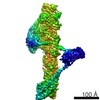
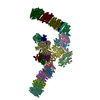
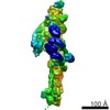
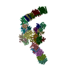
Journal: Cell / Year: 2021 Title: De novo identification of mammalian ciliary motility proteins using cryo-EM. Authors: Miao Gui / Hannah Farley / Priyanka Anujan / Jacob R Anderson / Dale W Maxwell / Jonathan B Whitchurch / J Josephine Botsch / Tao Qiu / Shimi Meleppattu / Sandeep K Singh / Qi Zhang / James ...Authors: Miao Gui / Hannah Farley / Priyanka Anujan / Jacob R Anderson / Dale W Maxwell / Jonathan B Whitchurch / J Josephine Botsch / Tao Qiu / Shimi Meleppattu / Sandeep K Singh / Qi Zhang / James Thompson / Jane S Lucas / Colin D Bingle / Dominic P Norris / Sudipto Roy / Alan Brown / Abstract: Dynein-decorated doublet microtubules (DMTs) are critical components of the oscillatory molecular machine of cilia, the axoneme, and have luminal surfaces patterned periodically by microtubule inner ...Dynein-decorated doublet microtubules (DMTs) are critical components of the oscillatory molecular machine of cilia, the axoneme, and have luminal surfaces patterned periodically by microtubule inner proteins (MIPs). Here we present an atomic model of the 48-nm repeat of a mammalian DMT, derived from a cryoelectron microscopy (cryo-EM) map of the complex isolated from bovine respiratory cilia. The structure uncovers principles of doublet microtubule organization and features specific to vertebrate cilia, including previously unknown MIPs, a luminal bundle of tektin filaments, and a pentameric dynein-docking complex. We identify a mechanism for bridging 48- to 24-nm periodicity across the microtubule wall and show that loss of the proteins involved causes defective ciliary motility and laterality abnormalities in zebrafish and mice. Our structure identifies candidate genes for diagnosis of ciliopathies and provides a framework to understand their functions in driving ciliary motility.
History
Deposition
Aug 10, 2021
-
Header (metadata) release
Oct 27, 2021
-
Map release
Oct 27, 2021
-
Update
Nov 24, 2021
-
Current status
Nov 24, 2021
Processing site: RCSB / Status: Released
-
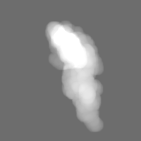
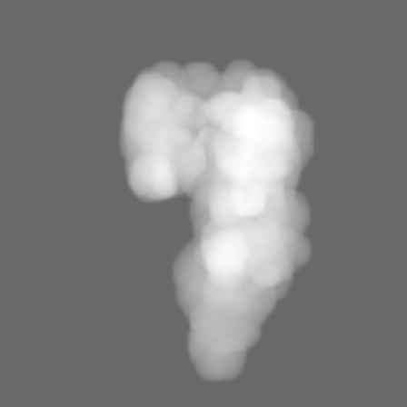
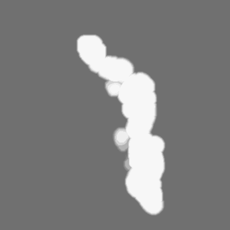
Structure visualization
Movie
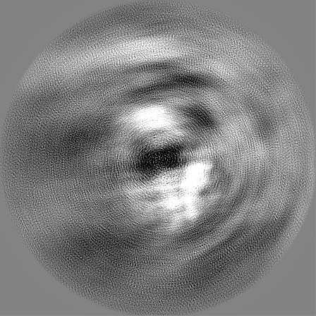
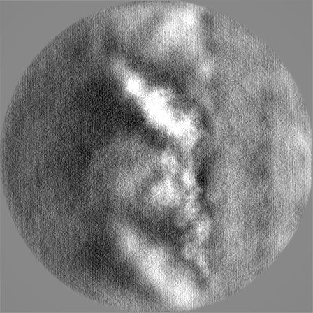
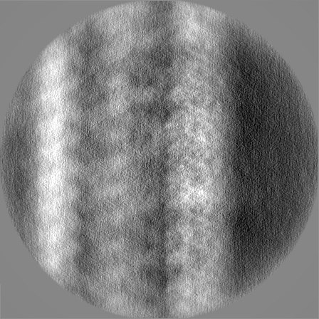
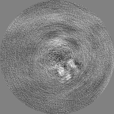
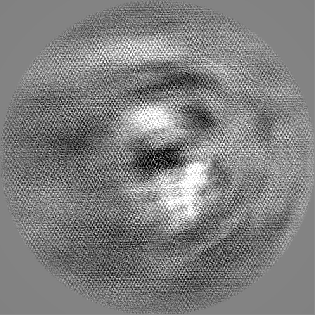
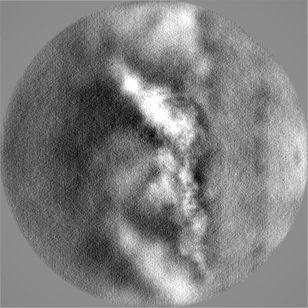
Surface view with section colored by density value
In the structure databanks used in Yorodumi, some data are registered as the other names, "COVID-19 virus" and "2019-nCoV". Here are the details of the virus and the list of structure data.
Jan 31, 2019. EMDB accession codes are about to change! (news from PDBe EMDB page)
EMDB accession codes are about to change! (news from PDBe EMDB page)
The allocation of 4 digits for EMDB accession codes will soon come to an end. Whilst these codes will remain in use, new EMDB accession codes will include an additional digit and will expand incrementally as the available range of codes is exhausted. The current 4-digit format prefixed with “EMD-” (i.e. EMD-XXXX) will advance to a 5-digit format (i.e. EMD-XXXXX), and so on. It is currently estimated that the 4-digit codes will be depleted around Spring 2019, at which point the 5-digit format will come into force.
The EM Navigator/Yorodumi systems omit the EMD- prefix.
Related info.:Q: What is EMD? / ID/Accession-code notation in Yorodumi/EM Navigator
Yorodumi is a browser for structure data from EMDB, PDB, SASBDB, etc.
This page is also the successor to EM Navigator detail page, and also detail information page/front-end page for Omokage search.
The word "yorodu" (or yorozu) is an old Japanese word meaning "ten thousand". "mi" (miru) is to see.
Related info.:EMDB / PDB / SASBDB / Comparison of 3 databanks / Yorodumi Search / Aug 31, 2016. New EM Navigator & Yorodumi / Yorodumi Papers / Jmol/JSmol / Function and homology information / Changes in new EM Navigator and Yorodumi
 Movie
Movie Controller

 Yorodumi
 Open data
 Basic information
 Map data
 Sample
 Authors
 United States, 1 items
  Citation

 Structure visualization
 Movie viewer
 Downloads & links
 emd_24663.png
 http://ftp.pdbj.org/pub/emdb/structures/EMD-24663


 Z (Sec.)
 Y (Row.)
 X (Col.)


 Sample components
 Processing
 Electron microscopy
 FIELD EMISSION GUN