ムービー
ムービー コントローラー


+ データを開く

- 基本情報
基本情報
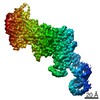
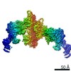
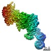
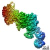
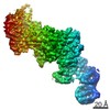
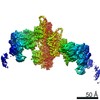
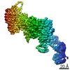
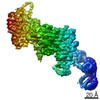
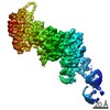
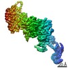
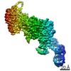
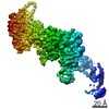
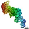
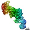
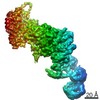
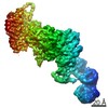
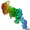
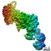
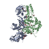
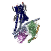
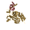
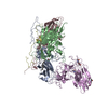
| 登録情報 | データベース: PDB / ID: 7pm3 | ||||||||||||||||||
|---|---|---|---|---|---|---|---|---|---|---|---|---|---|---|---|---|---|---|---|
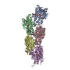
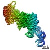
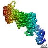
| タイトル | Cryo-EM structure of young JASP-stabilized F-actin (central 3er) | ||||||||||||||||||
 要素 要素 | Actin, alpha skeletal muscle | ||||||||||||||||||
 キーワード キーワード | STRUCTURAL PROTEIN / myosin / cytoskeleton / F-actin / jasplakinolide | ||||||||||||||||||
| 機能・相同性 |  機能・相同性情報 機能・相同性情報cytoskeletal motor activator activity / myosin heavy chain binding / tropomyosin binding / actin filament bundle / troponin I binding / filamentous actin / mesenchyme migration / actin filament bundle assembly / skeletal muscle myofibril / striated muscle thin filament ...cytoskeletal motor activator activity / myosin heavy chain binding / tropomyosin binding / actin filament bundle / troponin I binding / filamentous actin / mesenchyme migration / actin filament bundle assembly / skeletal muscle myofibril / striated muscle thin filament / skeletal muscle thin filament assembly / actin monomer binding / skeletal muscle fiber development / stress fiber / titin binding / actin filament polymerization / actin filament / filopodium / 加水分解酵素; 酸無水物に作用; 酸無水物に作用・細胞または細胞小器官の運動に関与 / calcium-dependent protein binding / lamellipodium / cell body / hydrolase activity / protein domain specific binding / calcium ion binding / positive regulation of gene expression / magnesium ion binding / ATP binding / identical protein binding / cytoplasm 類似検索 - 分子機能 | ||||||||||||||||||
| 生物種 |  | ||||||||||||||||||
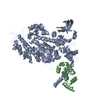
| 手法 | 電子顕微鏡法 / らせん対称体再構成法 / クライオ電子顕微鏡法 / 解像度: 3.1 Å | ||||||||||||||||||
 データ登録者 データ登録者 | Pospich, S. / Sweeney, H.L. / Houdusse, A. / Raunser, S. | ||||||||||||||||||
| 資金援助 |  ドイツ, European Union, ドイツ, European Union,  フランス, フランス,  米国, 5件 米国, 5件
| ||||||||||||||||||
 引用 引用 | ジャーナル: Elife / 年: 2021 タイトル: High-resolution structures of the actomyosin-V complex in three nucleotide states provide insights into the force generation mechanism. 著者: Sabrina Pospich / H Lee Sweeney / Anne Houdusse / Stefan Raunser / 要旨: The molecular motor myosin undergoes a series of major structural transitions during its force-producing motor cycle. The underlying mechanism and its coupling to ATP hydrolysis and actin binding are ...The molecular motor myosin undergoes a series of major structural transitions during its force-producing motor cycle. The underlying mechanism and its coupling to ATP hydrolysis and actin binding are only partially understood, mostly due to sparse structural data on actin-bound states of myosin. Here, we report 26 high-resolution cryo-EM structures of the actomyosin-V complex in the strong-ADP, rigor, and a previously unseen post-rigor transition state that binds the ATP analog AppNHp. The structures reveal a high flexibility of myosin in each state and provide valuable insights into the structural transitions of myosin-V upon ADP release and binding of AppNHp, as well as the actomyosin interface. In addition, they show how myosin is able to specifically alter the structure of F-actin. | ||||||||||||||||||
| 履歴 |
|
- 構造の表示
構造の表示
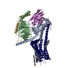
| ムービー |
ムービービューア |
|---|---|
| 構造ビューア | 分子: MolmilJmol/JSmol |
- ダウンロードとリンク
-ダウンロード
| PDBx/mmCIF形式 | 7pm3.cif.gz | 197.6 KB | 表示 | PDBx/mmCIF形式 |
|---|---|---|---|---|
| PDB形式 | pdb7pm3.ent.gz | 165.3 KB | 表示 | PDB形式 |
| PDBx/mmJSON形式 | 7pm3.json.gz | ツリー表示 | PDBx/mmJSON形式 | |
| その他 |  その他のダウンロード その他のダウンロード |
-検証レポート
| 文書・要旨 | 7pm3_validation.pdf.gz | 1.2 MB | 表示 | wwPDB検証レポート |
|---|---|---|---|---|
| 文書・詳細版 | 7pm3_full_validation.pdf.gz | 1.2 MB | 表示 | |
| XML形式データ | 7pm3_validation.xml.gz | 46.5 KB | 表示 | |
| CIF形式データ | 7pm3_validation.cif.gz | 63.3 KB | 表示 | |
| アーカイブディレクトリ | https://data.pdbj.org/pub/pdb/validation_reports/pm/7pm3ftp://data.pdbj.org/pub/pdb/validation_reports/pm/7pm3 | HTTPS FTP |
-関連構造データ
| 関連構造データ | 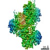 13511MC  7pltC 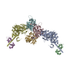 7pluC 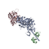 7plvC 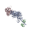 7plwC  7plxC 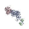 7plyC 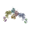 7plzC  7pm0C  7pm1C 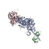 7pm2C 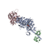 7pm5C 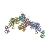 7pm6C 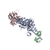 7pm7C 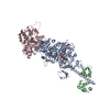 7pm8C  7pm9C 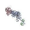 7pmaC 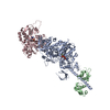 7pmbC 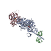 7pmcC  7pmdC  7pmeC 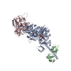 7pmfC 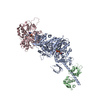 7pmgC 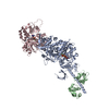 7pmhC 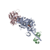 7pmiC 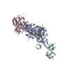 7pmjC  7pmlC C: 同じ文献を引用 ( M: このデータのモデリングに利用したマップデータ |
|---|---|
| 類似構造データ |


-リンク
 PDBj
PDBj


- 集合体
集合体
| 登録構造単位 | 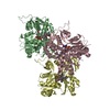
|
|---|---|
| 1 |
|
-要素
| #1: タンパク質 | 分子量: 42109.973 Da / 分子数: 3 / 由来タイプ: 天然 / 由来: (天然) #2: 化合物 |   分子量: 427.201 Da / 分子数: 3 / 由来タイプ: 合成 / 式: C10H15N5O10P2 / コメント: ADP, エネルギー貯蔵分子*YM 分子量: 427.201 Da / 分子数: 3 / 由来タイプ: 合成 / 式: C10H15N5O10P2 / コメント: ADP, エネルギー貯蔵分子*YM#3: 化合物 |   分子量: 94.971 Da / 分子数: 3 / 由来タイプ: 合成 / 式: PO4 分子量: 94.971 Da / 分子数: 3 / 由来タイプ: 合成 / 式: PO4#4: 化合物 |   分子量: 24.305 Da / 分子数: 3 / 由来タイプ: 合成 / 式: Mg 分子量: 24.305 Da / 分子数: 3 / 由来タイプ: 合成 / 式: Mg#5: 化合物 |   分子量: 709.670 Da / 分子数: 3 / 由来タイプ: 合成 / 式: C36H45BrN4O6 分子量: 709.670 Da / 分子数: 3 / 由来タイプ: 合成 / 式: C36H45BrN4O6研究の焦点であるリガンドがあるか | N | |
|---|
-実験情報
-実験
| 実験 | 手法: 電子顕微鏡法 |
|---|---|
| EM実験 | 試料の集合状態: FILAMENT / 3次元再構成法: らせん対称体再構成法 |
- 試料調製
試料調製
| 構成要素 | 名称: Young JASP-stabilized F-actin / タイプ: COMPLEX 詳細: Young ADP-Pi-bound F-actin stabilized with jasplakinolide Entity ID: #1 / 由来: NATURAL |
|---|---|
| 分子量 | 実験値: NO |
| 由来(天然) | 生物種: |
| 緩衝液 | pH: 7.5 |
| 試料 | 包埋: NO / シャドウイング: NO / 染色: NO / 凍結: YES / 詳細: Rise 27.9 A, Twist -166.9 degrees |
| 試料支持 | グリッドの材料: COPPER / グリッドのサイズ: 300 divisions/in. / グリッドのタイプ: Quantifoil R2/1 |
| 急速凍結 | 装置: FEI VITROBOT MARK III / 凍結剤: ETHANE / 湿度: 100 % / 凍結前の試料温度: 286 K |
- 電子顕微鏡撮影
| 実験機器 |  モデル: Titan Krios / 画像提供: FEI Company |
|---|---|
| 顕微鏡 | モデル: FEI TITAN KRIOS |
| 電子銃 | 電子線源:  FIELD EMISSION GUN / 加速電圧: 300 kV / 照射モード: SPOT SCAN FIELD EMISSION GUN / 加速電圧: 300 kV / 照射モード: SPOT SCAN |
| 電子レンズ | モード: BRIGHT FIELD / Cs: 0 mm |
| 試料ホルダ | 凍結剤: NITROGEN 試料ホルダーモデル: FEI TITAN KRIOS AUTOGRID HOLDER |
| 撮影 | 平均露光時間: 15 sec. / 電子線照射量: 80 e/Å2 / 検出モード: SUPER-RESOLUTION フィルム・検出器のモデル: GATAN K2 SUMMIT (4k x 4k) 撮影したグリッド数: 1 / 実像数: 1064 |
| 電子光学装置 | エネルギーフィルター名称: GIF Bioquantum / エネルギーフィルタースリット幅: 20 eV / 球面収差補正装置: Cs-corrected microscope |
| 画像スキャン | 動画フレーム数/画像: 40 |
- 解析
解析
| ソフトウェア | 名称: PHENIX / バージョン: 1.17.1_3660: / 分類: 精密化 | ||||||||||||||||||||||||||||||||||||
|---|---|---|---|---|---|---|---|---|---|---|---|---|---|---|---|---|---|---|---|---|---|---|---|---|---|---|---|---|---|---|---|---|---|---|---|---|---|
| EMソフトウェア |
| ||||||||||||||||||||||||||||||||||||
| CTF補正 | タイプ: PHASE FLIPPING AND AMPLITUDE CORRECTION | ||||||||||||||||||||||||||||||||||||
| 3次元再構成 | 解像度: 3.1 Å / 解像度の算出法: FSC 0.143 CUT-OFF / 粒子像の数: 212660 / 対称性のタイプ: POINT | ||||||||||||||||||||||||||||||||||||
| 原子モデル構築 | プロトコル: OTHER / 空間: REAL 詳細: An initial model of jasplakinolide was generated using elBow within Phenix inputting the SMILES string. | ||||||||||||||||||||||||||||||||||||
| 原子モデル構築 | PDB-ID: 5OOD PDB chain-ID: C | ||||||||||||||||||||||||||||||||||||
| 拘束条件 |
|