 Movie
Movie Controller

[English] 日本語
 Yorodumi
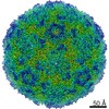
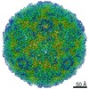
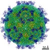
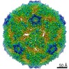
- PDB-7bzt: Cryo-EM structure of mature Coxsackievirus A10 in complex with KR... -
+ Open data

- Basic information
| Entry | Database: PDB / ID: 7bzt | ||||||||||||||||||||||||||||||||||||||||||||||||||||||
|---|---|---|---|---|---|---|---|---|---|---|---|---|---|---|---|---|---|---|---|---|---|---|---|---|---|---|---|---|---|---|---|---|---|---|---|---|---|---|---|---|---|---|---|---|---|---|---|---|---|---|---|---|---|---|---|
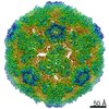
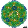
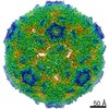
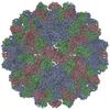
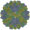
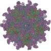
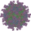
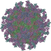
| Title | Cryo-EM structure of mature Coxsackievirus A10 in complex with KRM1 at pH 7.4 | ||||||||||||||||||||||||||||||||||||||||||||||||||||||
 Components Components |
| ||||||||||||||||||||||||||||||||||||||||||||||||||||||
 Keywords Keywords | VIRUS / Picornavirus / Coxsackievirus A10 / pH 7.4 / mature particle / KRM1 / complex | ||||||||||||||||||||||||||||||||||||||||||||||||||||||
| Function / homology |  Function and homology information Function and homology informationSignaling by LRP5 mutants / Negative regulation of TCF-dependent signaling by WNT ligand antagonists / cell communication / negative regulation of axon regeneration / negative regulation of ossification / regulation of canonical Wnt signaling pathway / limb development / symbiont-mediated suppression of host cytoplasmic pattern recognition receptor signaling pathway via inhibition of MDA-5 activity / picornain 2A / symbiont-mediated suppression of host mRNA export from nucleus ...Signaling by LRP5 mutants / Negative regulation of TCF-dependent signaling by WNT ligand antagonists / cell communication / negative regulation of axon regeneration / negative regulation of ossification / regulation of canonical Wnt signaling pathway / limb development / symbiont-mediated suppression of host cytoplasmic pattern recognition receptor signaling pathway via inhibition of MDA-5 activity / picornain 2A / symbiont-mediated suppression of host mRNA export from nucleus / TCF dependent signaling in response to WNT / symbiont genome entry into host cell via pore formation in plasma membrane / picornain 3C / T=pseudo3 icosahedral viral capsid / negative regulation of canonical Wnt signaling pathway / host cell cytoplasmic vesicle membrane / Wnt signaling pathway / ribonucleoside triphosphate phosphatase activity / transmembrane signaling receptor activity / nucleoside-triphosphate phosphatase / channel activity / monoatomic ion transmembrane transport / DNA replication / RNA helicase activity / endocytosis involved in viral entry into host cell / symbiont-mediated activation of host autophagy / RNA-directed RNA polymerase / cysteine-type endopeptidase activity / viral RNA genome replication / RNA-directed RNA polymerase activity / neuronal cell body / apoptotic process / virion attachment to host cell / DNA-templated transcription / host cell nucleus / structural molecule activity / signal transduction / proteolysis / RNA binding / zinc ion binding / ATP binding / membrane / plasma membrane Similarity search - Function | ||||||||||||||||||||||||||||||||||||||||||||||||||||||
| Biological species |  Homo sapiens (human) Homo sapiens (human) 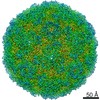 Coxsackievirus A10 Coxsackievirus A10 | ||||||||||||||||||||||||||||||||||||||||||||||||||||||
| Method | ELECTRON MICROSCOPY / single particle reconstruction / cryo EM / Resolution: 3 Å | ||||||||||||||||||||||||||||||||||||||||||||||||||||||
 Authors Authors | Cui, Y. / Peng, R. / Song, H. / Tong, Z. / Gao, G.F. / Qi, J. | ||||||||||||||||||||||||||||||||||||||||||||||||||||||
| Funding support |  China, 1items China, 1items
| ||||||||||||||||||||||||||||||||||||||||||||||||||||||
 Citation Citation | Journal: Proc Natl Acad Sci U S A / Year: 2020 Title: Molecular basis of Coxsackievirus A10 entry using the two-in-one attachment and uncoating receptor KRM1. Authors: Yingzi Cui / Ruchao Peng / Hao Song / Zhou Tong / Xiao Qu / Sheng Liu / Xin Zhao / Yan Chai / Peiyi Wang / George F Gao / Jianxun Qi / Abstract: KREMEN1 (KRM1) has been identified as a functional receptor for Coxsackievirus A10 (CV-A10), a causative agent of hand-foot-and-mouth disease (HFMD), which poses a great threat to infants globally. ...KREMEN1 (KRM1) has been identified as a functional receptor for Coxsackievirus A10 (CV-A10), a causative agent of hand-foot-and-mouth disease (HFMD), which poses a great threat to infants globally. However, the underlying mechanisms for the viral entry process are not well understood. Here we determined the atomic structures of different forms of CV-A10 viral particles and its complex with KRM1 in both neutral and acidic conditions. These structures reveal that KRM1 selectively binds to the mature viral particle above the canyon of the viral protein 1 (VP1) subunit and contacts across two adjacent asymmetry units. The key residues for receptor binding are conserved among most KRM1-dependent enteroviruses, suggesting a uniform mechanism for receptor binding. Moreover, the binding of KRM1 induces the release of pocket factor, a process accelerated under acidic conditions. Further biochemical studies confirmed that receptor binding at acidic pH enabled CV-A10 virion uncoating in vitro. Taken together, these findings provide high-resolution snapshots of CV-A10 entry and identify KRM1 as a two-in-one receptor for enterovirus infection. | ||||||||||||||||||||||||||||||||||||||||||||||||||||||
| History |
|
- Structure visualization
| Movie |
Movie viewer |
|---|---|
| Structure viewer | Molecule: MolmilJmol/JSmol |
- Downloads & links
-Download
| PDBx/mmCIF format | 7bzt.cif.gz | 209.4 KB | Display | PDBx/mmCIF format |
|---|---|---|---|---|
| PDB format | pdb7bzt.ent.gz | 163.3 KB | Display | PDB format |
| PDBx/mmJSON format | 7bzt.json.gz | Tree view | PDBx/mmJSON format | |
| Others |  Other downloads Other downloads |
-Validation report
| Arichive directory | https://data.pdbj.org/pub/pdb/validation_reports/bz/7bztftp://data.pdbj.org/pub/pdb/validation_reports/bz/7bzt | HTTPS FTP |
|---|
-Related structure data
| Related structure data | 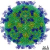 30259MC 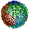 7bznC 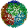 7bzoC 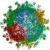 7bzuC 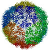 7c4tC  7c4wC 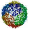 7c4yC 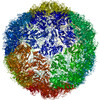 7c4zC M: map data used to model this data C: citing same article ( |
|---|---|
| Similar structure data |


-Links
 PDBj
PDBj


- Assembly
| Deposited unit | 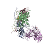
|
|---|---|
| 1 | x 60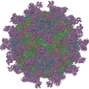
|
| 2 |
|
| 3 | x 5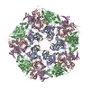
|
| 4 | x 6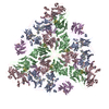
|
| 5 | 
|
| 6 | x 60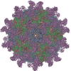
|
-Components
-Capsid protein ... , 4 types, 4 molecules ABCD
| #1: Protein | Mass: 33204.332 Da / Num. of mol.: 1 / Source method: isolated from a natural source / Source: (natural) Coxsackievirus A10 / References: UniProt: A0A0C5AWF6*PLUS |
|---|---|
| #2: Protein | Mass: 27783.105 Da / Num. of mol.: 1 / Source method: isolated from a natural source / Source: (natural) Coxsackievirus A10 / References: UniProt: G0YPI2 |
| #3: Protein | Mass: 26187.623 Da / Num. of mol.: 1 / Source method: isolated from a natural source / Source: (natural) Coxsackievirus A10 / References: UniProt: G0YPI2 |
| #4: Protein | Mass: 7464.104 Da / Num. of mol.: 1 / Source method: isolated from a natural source / Source: (natural) Coxsackievirus A10 / References: UniProt: G0YPI2 |
-Protein / Non-polymers / Sugars , 3 types, 5 molecules E

| #5: Protein | Mass: 41499.027 Da / Num. of mol.: 1 Source method: isolated from a genetically manipulated source Source: (gene. exp.) Homo sapiens (human) / Production host: Homo sapiens (human) / References: UniProt: Q96MU8*PLUS |
|---|---|
| #6: Chemical | ChemComp-SPH /  Mass: 299.492 Da / Num. of mol.: 1 / Source method: obtained synthetically / Formula: C18H37NO2 / Feature type: SUBJECT OF INVESTIGATION Mass: 299.492 Da / Num. of mol.: 1 / Source method: obtained synthetically / Formula: C18H37NO2 / Feature type: SUBJECT OF INVESTIGATION |
| #7: Sugar |  Type: D-saccharide, beta linking / Mass: 221.208 Da / Num. of mol.: 3 Type: D-saccharide, beta linking / Mass: 221.208 Da / Num. of mol.: 3Source method: isolated from a genetically manipulated source Formula: C8H15NO6 |
-Details
| Has ligand of interest | Y |
|---|---|
| Has protein modification | Y |
| Sequence details | The genome sequence of chain A have been deposit in GenBank: MT263729 |
-Experimental details
-Experiment
| Experiment | Method: ELECTRON MICROSCOPY |
|---|---|
| EM experiment | Aggregation state: PARTICLE / 3D reconstruction method: single particle reconstruction |
- Sample preparation
| Component | Name: Coxsackievirus A10 / Type: VIRUS / Entity ID: #1-#5 / Source: NATURAL |
|---|---|
| Source (natural) | Organism: Coxsackievirus A10 |
| Details of virus | Empty: NO / Enveloped: NO / Isolate: STRAIN / Type: VIRION |
| Buffer solution | pH: 7.4 |
| Specimen | Embedding applied: NO / Shadowing applied: NO / Staining applied: NO / Vitrification applied: YES |
| Vitrification | Instrument: FEI VITROBOT MARK IV / Cryogen name: ETHANE / Humidity: 100 % / Chamber temperature: 277 K |
- Electron microscopy imaging
| Experimental equipment |  Model: Talos Arctica / Image courtesy: FEI Company | ||||||||||||||||
|---|---|---|---|---|---|---|---|---|---|---|---|---|---|---|---|---|---|
| Microscopy | Model: FEI TALOS ARCTICA | ||||||||||||||||
| Electron gun | Electron source:  FIELD EMISSION GUN / Accelerating voltage: 200 kV / Illumination mode: FLOOD BEAM FIELD EMISSION GUN / Accelerating voltage: 200 kV / Illumination mode: FLOOD BEAM | ||||||||||||||||
| Electron lens | Mode: BRIGHT FIELD | ||||||||||||||||
| Image recording |
|
- Processing
| Software | Name: PHENIX / Version: 1.17.1_3660: / Classification: refinement | ||||||||||||||||||||||||
|---|---|---|---|---|---|---|---|---|---|---|---|---|---|---|---|---|---|---|---|---|---|---|---|---|---|
| EM software | Name: PHENIX / Category: model refinement | ||||||||||||||||||||||||
| CTF correction | Type: PHASE FLIPPING AND AMPLITUDE CORRECTION | ||||||||||||||||||||||||
| 3D reconstruction | Resolution: 3 Å / Resolution method: FSC 0.143 CUT-OFF / Num. of particles: 28084 / Symmetry type: POINT | ||||||||||||||||||||||||
| Refine LS restraints |
|
