#262 - Oct 2021 Fifty Years of Open Access to PDB Structures similarity (3)
#200 - Aug 2016 Quasisymmetry in Icosahedral Viruses similarity (4)
-
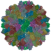
Assembly
Deposited unit
A: Capsid protein B: Capsid protein D: Capsid protein E: Capsid protein F: Capsid protein G: Capsid protein H: Capsid protein I: Capsid protein J: Capsid protein K: Capsid protein L: Capsid protein M: Capsid protein N: Capsid protein O: Capsid protein P: Capsid protein hetero molecules
A: Capsid protein B: Capsid protein D: Capsid protein E: Capsid protein F: Capsid protein G: Capsid protein H: Capsid protein I: Capsid protein J: Capsid protein K: Capsid protein L: Capsid protein M: Capsid protein N: Capsid protein O: Capsid protein P: Capsid protein hetero molecules
Idetical with deposited unit in distinct coordinate
point asymmetric unit, std point frame
Type
Name
Symmetry operation
Number
transform to point frame
1
4
A: Capsid protein B: Capsid protein D: Capsid protein E: Capsid protein F: Capsid protein G: Capsid protein H: Capsid protein I: Capsid protein J: Capsid protein K: Capsid protein L: Capsid protein M: Capsid protein N: Capsid protein O: Capsid protein P: Capsid protein hetero molecules
crystal asymmetric unit, crystal frame
615 kDa, 15 polymers
Theoretical mass
Number of molelcules
Total (without water)
614,639
35
Polymers
613,711
15
Non-polymers
928
20
Water
270
15
Type
Name
Symmetry operation
Number
identity operation
1_555
x,y,z
2
Unit cell
Length a, b, c (Å)
384.000, 384.000, 384.000
Angle α, β, γ (deg.)
90.00, 90.00, 90.00
Int Tables number
197
Space group name H-M
I23
Symmetry
Point symmetry: (Schoenflies symbol: I (icosahedral))
Components on special symmetry positions
ID
Model
Components
1
1
G-401-
ZN
2
1
M-401-
ZN
3
1
G-503-
HOH
Noncrystallographic symmetry (NCS)
NCS domain:
ID
Ens-ID
1
1
2
1
3
1
4
1
5
1
1
2
2
2
3
2
4
2
5
2
1
3
2
3
3
3
4
3
5
3
NCS domain segments:
Dom-ID
Component-ID
Ens-ID
Selection details
1
1
1
ChainA
2
1
1
ChainE
3
1
1
chainH
4
1
1
chainK
5
1
1
chainN
1
1
2
ChainB
2
1
2
chainF
3
1
2
chainI
4
1
2
chainL
5
1
2
ChainO
1
1
3
chainD
2
1
3
chainG
3
1
3
chainJ
4
1
3
chainM
5
1
3
chainP
NCS ensembles :
ID
1
2
3
-
Components
#1: Protein
Capsidprotein / Coat protein / p41
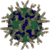
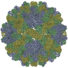
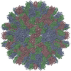
Mass: 40914.043 Da / Num. of mol.: 15 / Source method: isolated from a natural source / Source: (natural) Cucumber necrosis virus / Strain: Nicotiana benthamiana / References: UniProt: P15183
Mass: 18.015 Da / Num. of mol.: 61 / Source method: isolated from a natural source / Formula: H2O
-
Experimental details
-
Experiment
Experiment
Method: X-RAY DIFFRACTION / Number of used crystals: 4
-
Sample preparation
Crystal
Density Matthews: 3.84 Å3/Da / Density % sol: 68 %
Crystal grow
Temperature: 295 K / Method: vapor diffusion, hanging drop / pH: 6 Details: Virus concentration was adjusted to 10 mg/ml in 20 mM sodium acetate buffer at pH 5.0. The reservoir contained 0.16 M PIPES buffer and 3.2 M sodium formate, pH 6.0. Drop composed of 10 ...Details: Virus concentration was adjusted to 10 mg/ml in 20 mM sodium acetate buffer at pH 5.0. The reservoir contained 0.16 M PIPES buffer and 3.2 M sodium formate, pH 6.0. Drop composed of 10 microliters reservoir and virus solutions., VAPOR DIFFUSION, HANGING DROP, temperature 295K
In the structure databanks used in Yorodumi, some data are registered as the other names, "COVID-19 virus" and "2019-nCoV". Here are the details of the virus and the list of structure data.
Jan 31, 2019. EMDB accession codes are about to change! (news from PDBe EMDB page)
EMDB accession codes are about to change! (news from PDBe EMDB page)
The allocation of 4 digits for EMDB accession codes will soon come to an end. Whilst these codes will remain in use, new EMDB accession codes will include an additional digit and will expand incrementally as the available range of codes is exhausted. The current 4-digit format prefixed with “EMD-” (i.e. EMD-XXXX) will advance to a 5-digit format (i.e. EMD-XXXXX), and so on. It is currently estimated that the 4-digit codes will be depleted around Spring 2019, at which point the 5-digit format will come into force.
The EM Navigator/Yorodumi systems omit the EMD- prefix.
Related info.:Q: What is EMD? / ID/Accession-code notation in Yorodumi/EM Navigator
Yorodumi is a browser for structure data from EMDB, PDB, SASBDB, etc.
This page is also the successor to EM Navigator detail page, and also detail information page/front-end page for Omokage search.
The word "yorodu" (or yorozu) is an old Japanese word meaning "ten thousand". "mi" (miru) is to see.
Related info.:EMDB / PDB / SASBDB / Comparison of 3 databanks / Yorodumi Search / Aug 31, 2016. New EM Navigator & Yorodumi / Yorodumi Papers / Jmol/JSmol / Function and homology information / Changes in new EM Navigator and Yorodumi
 Movie
Movie Controller


 Open data
 Basic information
 Components
 Keywords
 Function and homology information
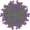
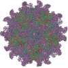
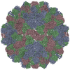
 Cucumber necrosis virus
 X-RAY DIFFRACTION /
  Authors
 Citation
 Structure visualization
 Downloads & links
 Other downloads
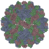


 PDBj
PDBj

 Assembly
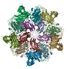


 Mass: 40.078 Da / Num. of mol.: 15 / Source method: obtained synthetically / Formula: Ca
 Mass: 65.409 Da / Num. of mol.: 5 / Source method: obtained synthetically / Formula: Zn
 Mass: 18.015 Da / Num. of mol.: 61 / Source method: isolated from a natural source / Formula: H2O
 Sample preparation
 Processing