 Movie
Movie Controller

[English] 日本語
 Yorodumi
- PDB-7p1i: Cryo EM structure of bison NHA2 in detergent and N-terminal exten... -
+ Open data

- Basic information
| Entry | Database: PDB / ID: 7p1i | |||||||||||||||||||||||||||||||||||||||||||||||||||||||||||||||||||||
|---|---|---|---|---|---|---|---|---|---|---|---|---|---|---|---|---|---|---|---|---|---|---|---|---|---|---|---|---|---|---|---|---|---|---|---|---|---|---|---|---|---|---|---|---|---|---|---|---|---|---|---|---|---|---|---|---|---|---|---|---|---|---|---|---|---|---|---|---|---|---|
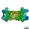
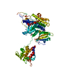
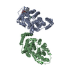
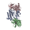
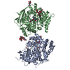
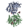
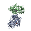
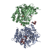
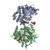
| Title | Cryo EM structure of bison NHA2 in detergent and N-terminal extension helix | |||||||||||||||||||||||||||||||||||||||||||||||||||||||||||||||||||||
 Components Components | mitochondrial sodium/hydrogen exchanger 9B2 | |||||||||||||||||||||||||||||||||||||||||||||||||||||||||||||||||||||
 Keywords Keywords | TRANSPORT PROTEIN / Membrane protein Sodium proton transporter | |||||||||||||||||||||||||||||||||||||||||||||||||||||||||||||||||||||
| Function / homology |  Function and homology information Function and homology informationlithium:proton antiporter activity / lithium ion transport / positive regulation of osteoclast development / sodium ion homeostasis / sodium:proton antiporter activity / regulation of insulin secretion involved in cellular response to glucose stimulus / clathrin-dependent endocytosis / flagellated sperm motility / sodium ion transport / recycling endosome ...lithium:proton antiporter activity / lithium ion transport / positive regulation of osteoclast development / sodium ion homeostasis / sodium:proton antiporter activity / regulation of insulin secretion involved in cellular response to glucose stimulus / clathrin-dependent endocytosis / flagellated sperm motility / sodium ion transport / recycling endosome / mitochondrial membrane / recycling endosome membrane / synaptic vesicle membrane / sperm principal piece / basolateral plasma membrane / endosome membrane / apical plasma membrane / lysosomal membrane / metal ion binding / identical protein binding / plasma membrane Similarity search - Function | |||||||||||||||||||||||||||||||||||||||||||||||||||||||||||||||||||||
| Biological species |  Bison bison (American bison) Bison bison (American bison) | |||||||||||||||||||||||||||||||||||||||||||||||||||||||||||||||||||||
| Method | ELECTRON MICROSCOPY / single particle reconstruction / cryo EM / Resolution: 3.15 Å | |||||||||||||||||||||||||||||||||||||||||||||||||||||||||||||||||||||
 Authors Authors | Matsuoka, R. / Fudim, R. / Jung, S. / Drew, D. | |||||||||||||||||||||||||||||||||||||||||||||||||||||||||||||||||||||
| Funding support | 1items
| |||||||||||||||||||||||||||||||||||||||||||||||||||||||||||||||||||||
 Citation Citation | Journal: Nat Struct Mol Biol / Year: 2022 Title: Structure, mechanism and lipid-mediated remodeling of the mammalian Na/H exchanger NHA2. Authors: Rei Matsuoka / Roman Fudim / Sukkyeong Jung / Chenou Zhang / Andre Bazzone / Yurie Chatzikyriakidou / Carol V Robinson / Norimichi Nomura / So Iwata / Michael Landreh / Laura Orellana / ...Authors: Rei Matsuoka / Roman Fudim / Sukkyeong Jung / Chenou Zhang / Andre Bazzone / Yurie Chatzikyriakidou / Carol V Robinson / Norimichi Nomura / So Iwata / Michael Landreh / Laura Orellana / Oliver Beckstein / David Drew /      Abstract: The Na/H exchanger SLC9B2, also known as NHA2, correlates with the long-sought-after Na/Li exchanger linked to the pathogenesis of diabetes mellitus and essential hypertension in humans. Despite the ...The Na/H exchanger SLC9B2, also known as NHA2, correlates with the long-sought-after Na/Li exchanger linked to the pathogenesis of diabetes mellitus and essential hypertension in humans. Despite the functional importance of NHA2, structural information and the molecular basis for its ion-exchange mechanism have been lacking. Here we report the cryo-EM structures of bison NHA2 in detergent and in nanodiscs, at 3.0 and 3.5 Å resolution, respectively. The bison NHA2 structure, together with solid-state membrane-based electrophysiology, establishes the molecular basis for electroneutral ion exchange. NHA2 consists of 14 transmembrane (TM) segments, rather than the 13 TMs previously observed in mammalian Na/H exchangers (NHEs) and related bacterial antiporters. The additional N-terminal helix in NHA2 forms a unique homodimer interface with a large intracellular gap between the protomers, which closes in the presence of phosphoinositol lipids. We propose that the additional N-terminal helix has evolved as a lipid-mediated remodeling switch for the regulation of NHA2 activity. #1: Journal: Nat.Struct.Mol.Biol. / Year: 2022Title: Structure, mechanism and lipid-mediated remodeling of the mammalian Na+/H+ exchanger NHA2 Authors: Matsuoka, R. / Fudim, F. / Jung, S. / Drew, F. | |||||||||||||||||||||||||||||||||||||||||||||||||||||||||||||||||||||
| History |
|
- Structure visualization
| Movie |
Movie viewer |
|---|---|
| Structure viewer | Molecule: MolmilJmol/JSmol |
- Downloads & links
-Download
| PDBx/mmCIF format | 7p1i.cif.gz | 155.4 KB | Display | PDBx/mmCIF format |
|---|---|---|---|---|
| PDB format | pdb7p1i.ent.gz | 121.8 KB | Display | PDB format |
| PDBx/mmJSON format | 7p1i.json.gz | Tree view | PDBx/mmJSON format | |
| Others |  Other downloads Other downloads |
-Validation report
| Arichive directory | https://data.pdbj.org/pub/pdb/validation_reports/p1/7p1iftp://data.pdbj.org/pub/pdb/validation_reports/p1/7p1i | HTTPS FTP |
|---|
-Related structure data
| Related structure data | 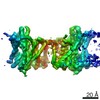 13161MC 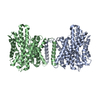 7p1jC  7p1kC M: map data used to model this data C: citing same article ( |
|---|---|
| Similar structure data |


-Links
 PDBj
PDBj- Assembly
| Deposited unit | 
|
|---|---|
| 1 |
|
-Components
| #1: Protein | Mass: 57419.203 Da / Num. of mol.: 2 Source method: isolated from a genetically manipulated source Source: (gene. exp.) Bison bison (American bison) / Production host:  Has protein modification | N | |
|---|
-Experimental details
-Experiment
| Experiment | Method: ELECTRON MICROSCOPY |
|---|---|
| EM experiment | Aggregation state: PARTICLE / 3D reconstruction method: single particle reconstruction |
- Sample preparation
| Component | Name: detergent structure / Type: COMPLEX / Entity ID: all / Source: RECOMBINANT | ||||||||||||||||||||
|---|---|---|---|---|---|---|---|---|---|---|---|---|---|---|---|---|---|---|---|---|---|
| Molecular weight | Value: 57373 kDa/nm / Experimental value: YES | ||||||||||||||||||||
| Source (natural) | Organism: Bison bison (American bison) | ||||||||||||||||||||
| Source (recombinant) | Organism: | ||||||||||||||||||||
| Buffer solution | pH: 7 | ||||||||||||||||||||
| Buffer component |
| ||||||||||||||||||||
| Specimen | Conc.: 5 mg/ml / Embedding applied: NO / Shadowing applied: NO / Staining applied: NO / Vitrification applied: YES | ||||||||||||||||||||
| Specimen support | Grid material: COPPER / Grid mesh size: 300 divisions/in. / Grid type: Quantifoil R2/1 | ||||||||||||||||||||
| Vitrification | Instrument: FEI VITROBOT MARK II / Cryogen name: ETHANE / Humidity: 100 % / Chamber temperature: 277 K |
- Electron microscopy imaging
| Experimental equipment |  Model: Titan Krios / Image courtesy: FEI Company |
|---|---|
| Microscopy | Model: FEI TITAN KRIOS |
| Electron gun | Electron source:  FIELD EMISSION GUN / Accelerating voltage: 300 kV / Illumination mode: FLOOD BEAM FIELD EMISSION GUN / Accelerating voltage: 300 kV / Illumination mode: FLOOD BEAM |
| Electron lens | Mode: BRIGHT FIELD |
| Image recording | Electron dose: 80 e/Å2 / Detector mode: COUNTING / Film or detector model: GATAN K2 QUANTUM (4k x 4k) |
| Image scans | Width: 3838 / Height: 3710 |
- Processing
| Software | Name: PHENIX / Version: 1.19_4092: / Classification: refinement | ||||||||||||||||||||||||||||
|---|---|---|---|---|---|---|---|---|---|---|---|---|---|---|---|---|---|---|---|---|---|---|---|---|---|---|---|---|---|
| EM software |
| ||||||||||||||||||||||||||||
| CTF correction | Type: PHASE FLIPPING AND AMPLITUDE CORRECTION | ||||||||||||||||||||||||||||
| 3D reconstruction | Resolution: 3.15 Å / Resolution method: FSC 0.143 CUT-OFF / Num. of particles: 255710 / Algorithm: FOURIER SPACE / Symmetry type: POINT | ||||||||||||||||||||||||||||
| Refine LS restraints |
|
