Protocol: SINGLE WAVELENGTH / Monochromatic (M) / Laue (L): M / Scattering type: x-ray
Radiation wavelength
Wavelength: 1 Å / Relative weight: 1
Reflection
Resolution: 2.802→44.216 Å / Num. obs: 43693 / % possible obs: 99.6 % / Redundancy: 6.8 % / Rmerge(I) obs: 0.139 / Net I/σ(I): 13.03
Reflection shell
Resolution: 2.802→2.902 Å
-
Processing
Software
Name
Version
Classification
PHENIX
(1.12_2829: ???)
refinement
XDS
datareduction
XSCALE
datascaling
PHENIX
phasing
Refinement
Method to determine structure: MOLECULAR REPLACEMENT Starting model: It was solved by MR on a crude model from experimental phasing experiment (data not deposited) Resolution: 2.802→44.216 Å / SU ML: 0.49 / Cross valid method: FREE R-VALUE / σ(F): 1.2 / Phase error: 35.61
Rfactor
Num. reflection
% reflection
Rfree
0.2932
2199
5.03 %
Rwork
0.2578
-
-
obs
0.2597
43693
99.17 %
Solvent computation
Shrinkage radii: 0.9 Å / VDW probe radii: 1.11 Å
Refinement step
Cycle: LAST / Resolution: 2.802→44.216 Å
Protein
Nucleic acid
Ligand
Solvent
Total
Num. atoms
4838
0
25
0
4863
Refine LS restraints
Refine-ID
Type
Dev ideal
Number
X-RAY DIFFRACTION
f_bond_d
0.003
4957
X-RAY DIFFRACTION
f_angle_d
0.505
6717
X-RAY DIFFRACTION
f_dihedral_angle_d
14.518
2958
X-RAY DIFFRACTION
f_chiral_restr
0.038
751
X-RAY DIFFRACTION
f_plane_restr
0.003
880
LS refinement shell
Resolution (Å)
Rfactor Rfree
Num. reflection Rfree
Rfactor Rwork
Num. reflection Rwork
Refine-ID
% reflection obs (%)
2.8018-2.8627
0.4155
127
0.4571
2351
X-RAY DIFFRACTION
92
2.8627-2.9293
0.3863
138
0.4056
2610
X-RAY DIFFRACTION
100
2.9293-3.0025
0.386
141
0.3849
2618
X-RAY DIFFRACTION
99
3.0025-3.0837
0.3822
141
0.3622
2639
X-RAY DIFFRACTION
100
3.0837-3.1744
0.3519
136
0.3577
2581
X-RAY DIFFRACTION
100
3.1744-3.2769
0.4075
136
0.3395
2619
X-RAY DIFFRACTION
100
3.2769-3.3939
0.3472
138
0.3023
2634
X-RAY DIFFRACTION
100
3.3939-3.5298
0.3399
134
0.2852
2596
X-RAY DIFFRACTION
100
3.5298-3.6903
0.3472
141
0.2856
2623
X-RAY DIFFRACTION
100
3.6903-3.8848
0.3024
137
0.262
2579
X-RAY DIFFRACTION
100
3.8848-4.128
0.276
139
0.2419
2626
X-RAY DIFFRACTION
100
4.128-4.4465
0.2696
136
0.2226
2591
X-RAY DIFFRACTION
100
4.4465-4.8934
0.2247
137
0.2015
2604
X-RAY DIFFRACTION
100
4.8934-5.6003
0.2415
139
0.2153
2621
X-RAY DIFFRACTION
100
5.6003-7.0512
0.3156
141
0.2399
2618
X-RAY DIFFRACTION
100
7.0512-44.2217
0.2246
138
0.1962
2584
X-RAY DIFFRACTION
99
Refinement TLS params.
Method: refined / Refine-ID: X-RAY DIFFRACTION
ID
L11 (°2)
L12 (°2)
L13 (°2)
L22 (°2)
L23 (°2)
L33 (°2)
S11 (Å °)
S12 (Å °)
S13 (Å °)
S21 (Å °)
S22 (Å °)
S23 (Å °)
S31 (Å °)
S32 (Å °)
S33 (Å °)
T11 (Å2)
T12 (Å2)
T13 (Å2)
T22 (Å2)
T23 (Å2)
T33 (Å2)
Origin x (Å)
Origin y (Å)
Origin z (Å)
1
1.9243
0.1906
-0.8122
3.0586
-0.4504
4.1816
-0.1068
-0.0299
-0.0732
-0.2487
0.0102
-0.2703
0.4007
0.1552
0.0721
0.5986
-0.0356
-0.1105
0.3132
-0.0455
0.5004
-35.8783
-42.4299
4.3208
2
3.665
0.7351
1.2839
6.1579
1.2225
4.8527
-0.1318
0.3287
-0.4097
-0.5817
-0.2215
0.5643
0.5368
-0.8095
0.2959
0.5696
-0.1304
-0.0277
0.5513
-0.0773
0.4698
-55.7819
-44.2913
3.662
3
2.2827
0.3609
0.1222
3.6112
0.1303
2.6525
-0.0023
0.0861
-0.0235
-0.1517
-0.1908
0.6133
0.1589
-0.6496
0.1956
0.6191
0.0575
-0.1175
0.6439
-0.052
0.496
-58.1865
-42.9564
-1.31
4
2.1024
-1.4862
0.307
1.0557
-0.2501
0.4342
-0.2561
0.238
0.1033
-0.2768
0.234
0.2443
-0.4138
0.0414
-0.0074
1.3979
0.0009
-0.3278
1.0185
0.0337
0.7397
-74.0043
-2.7431
16.9704
5
5.3628
0.9649
1.9344
5.6582
0.3423
6.6198
-0.2064
0.3701
0.5138
0.1587
-0.1107
-0.5163
-0.8589
0.6379
0.3069
0.8462
-0.0747
-0.1558
0.6914
0.1236
0.4948
-66.1539
8.8385
24.6663
6
3.5623
1.654
1.166
5.4894
2.3692
4.8271
-0.3558
-0.6211
0.2076
0.381
0.1563
-0.2053
-0.1448
-0.4647
0.1008
0.847
0.1434
-0.0466
0.7199
-0.0291
0.5139
-48.6005
-38.0601
30.9949
7
1.0316
0.539
2.3854
2.5998
3.1409
7.2316
-1.1095
0.3918
0.3153
-0.2309
0.4305
0.2903
-0.3445
0.4773
0.7846
1.1094
0.1009
-0.2275
0.7764
-0.0168
0.5588
-42.8811
-26.4828
35.226
8
8.5583
1.2165
4.8608
5.9669
1.3637
5.6647
-0.575
-0.7543
0.9613
0.6334
0.2631
-0.0056
-0.557
-0.2789
0.3651
1.0119
0.1182
-0.1455
0.6923
-0.0726
0.4634
-45.6291
-31.374
34.126
Refinement TLS group
ID
Refine-ID
Refine TLS-ID
Selection details
1
X-RAY DIFFRACTION
1
(chainAandresid222:413)
2
X-RAY DIFFRACTION
2
(chainAandresid414:465)
3
X-RAY DIFFRACTION
3
(chainAandresid466:582)
4
X-RAY DIFFRACTION
4
(chainAandresid583:632)
5
X-RAY DIFFRACTION
5
(chainAandresid633:756)
6
X-RAY DIFFRACTION
6
(chainAandresid757:795)
7
X-RAY DIFFRACTION
7
(chainAandresid796:829)
8
X-RAY DIFFRACTION
8
(chainAandresid830:901)
+
About Yorodumi
-
News
-
Feb 9, 2022. New format data for meta-information of EMDB entries
New format data for meta-information of EMDB entries
Version 3 of the EMDB header file is now the official format.
The previous official version 1.9 will be removed from the archive.
In the structure databanks used in Yorodumi, some data are registered as the other names, "COVID-19 virus" and "2019-nCoV". Here are the details of the virus and the list of structure data.
Jan 31, 2019. EMDB accession codes are about to change! (news from PDBe EMDB page)
EMDB accession codes are about to change! (news from PDBe EMDB page)
The allocation of 4 digits for EMDB accession codes will soon come to an end. Whilst these codes will remain in use, new EMDB accession codes will include an additional digit and will expand incrementally as the available range of codes is exhausted. The current 4-digit format prefixed with “EMD-” (i.e. EMD-XXXX) will advance to a 5-digit format (i.e. EMD-XXXXX), and so on. It is currently estimated that the 4-digit codes will be depleted around Spring 2019, at which point the 5-digit format will come into force.
The EM Navigator/Yorodumi systems omit the EMD- prefix.
Related info.:Q: What is EMD? / ID/Accession-code notation in Yorodumi/EM Navigator
Yorodumi is a browser for structure data from EMDB, PDB, SASBDB, etc.
This page is also the successor to EM Navigator detail page, and also detail information page/front-end page for Omokage search.
The word "yorodu" (or yorozu) is an old Japanese word meaning "ten thousand". "mi" (miru) is to see.
Related info.:EMDB / PDB / SASBDB / Comparison of 3 databanks / Yorodumi Search / Aug 31, 2016. New EM Navigator & Yorodumi / Yorodumi Papers / Jmol/JSmol / Function and homology information / Changes in new EM Navigator and Yorodumi
 Movie
Movie Controller


 Open data
 Basic information
 Components
 Keywords
 Function and homology information
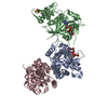
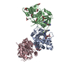
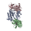
 Legionella pneumophila (bacteria)
 X-RAY DIFFRACTION /
  Authors
 Germany, 3items
  Citation
 Structure visualization
 Downloads & links
 Other downloads


 PDBj
PDBj Assembly
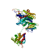
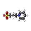
 Mass: 201.243 Da / Num. of mol.: 1 / Source method: obtained synthetically / Formula: C8H11NO3S
 Mass: 62.068 Da / Num. of mol.: 3 / Source method: obtained synthetically / Formula: C2H6O2
 Sample preparation
 / Beamline: X06DA / Wavelength: 1 Å
 Processing