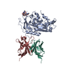
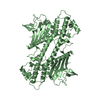
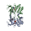
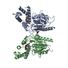
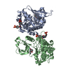
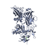
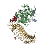
Chicken Scap D435V L1-L7 domain / Fab complex focused map
Components
4G10 Fab heavy chain
4G10 Fab kappa chain
Sterol regulatory element-binding protein cleavage-activating protein
Keywords
LIPID BINDING PROTEIN / Cholesterol
Function / homology
Function and homology information
Regulation of cholesterol biosynthesis by SREBP (SREBF) / SREBP-SCAP complex / regulation of cholesterol biosynthetic process / sterol binding / SREBP signaling pathway / regulation of fatty acid biosynthetic process / cholesterol metabolic process / ER to Golgi transport vesicle membrane / positive regulation of cholesterol biosynthetic process / response to insulin ...Regulation of cholesterol biosynthesis by SREBP (SREBF) / SREBP-SCAP complex / regulation of cholesterol biosynthetic process / sterol binding / SREBP signaling pathway / regulation of fatty acid biosynthetic process / cholesterol metabolic process / ER to Golgi transport vesicle membrane / positive regulation of cholesterol biosynthetic process / response to insulin / response to hypoxia / immune response / Golgi membrane / endoplasmic reticulum membrane Similarity search - Function
National Institutes of Health/National Institute of General Medical Sciences (NIH/NIGMS)
R35GM116387
United States
National Institutes of Health/National Heart, Lung, and Blood Institute (NIH/NHLBI)
P01HL020948
United States
National Institutes of Health/National Institute of General Medical Sciences (NIH/NIGMS)
R01GM136976
United States
Welch Foundation
I-1770
United States
Welch Foundation
I-1793
United States
Welch Foundation
I-1944
United States
Mallinckrodt Foundation
United States
Leducq Foundation
19CVD04
France
Cancer Prevention and Research Institute of Texas (CPRIT)
RR160082
United States
Cancer Prevention and Research Institute of Texas (CPRIT)
RP170644
United States
American Heart Association
18POST34080141
United States
Citation
Journal: Cell / Year: 2021 Title: Scap structures highlight key role for rotation of intertwined luminal loops in cholesterol sensing. Authors: Daniel L Kober / Arun Radhakrishnan / Joseph L Goldstein / Michael S Brown / Lindsay D Clark / Xiao-Chen Bai / Daniel M Rosenbaum / Abstract: The cholesterol-sensing protein Scap induces cholesterol synthesis by transporting membrane-bound transcription factors called sterol regulatory element-binding proteins (SREBPs) from the endoplasmic ...The cholesterol-sensing protein Scap induces cholesterol synthesis by transporting membrane-bound transcription factors called sterol regulatory element-binding proteins (SREBPs) from the endoplasmic reticulum (ER) to the Golgi apparatus for proteolytic activation. Transport requires interaction between Scap's two ER luminal loops (L1 and L7), which flank an intramembrane sterol-sensing domain (SSD). Cholesterol inhibits Scap transport by binding to L1, which triggers Scap's binding to Insig, an ER retention protein. Here we used cryoelectron microscopy (cryo-EM) to elucidate two structures of full-length chicken Scap: (1) a wild-type free of Insigs and (2) mutant Scap bound to chicken Insig without cholesterol. Strikingly, L1 and L7 intertwine tightly to form a globular domain that acts as a luminal platform connecting the SSD to the rest of Scap. In the presence of Insig, this platform undergoes a large rotation accompanied by rearrangement of Scap's transmembrane helices. We postulate that this conformational change halts Scap transport of SREBPs and inhibits cholesterol synthesis.
History
Deposition
Feb 2, 2021
Deposition site: RCSB / Processing site: RCSB
Revision 1.0
Jun 30, 2021
Provider: repository / Type: Initial release
Revision 1.0
Jun 30, 2021
Data content type: EM metadata / Data content type: EM metadata / Provider: repository / Type: Initial release
Revision 1.0
Jun 30, 2021
Data content type: FSC / Data content type: FSC / Provider: repository / Type: Initial release
Revision 1.0
Jun 30, 2021
Data content type: Image / Data content type: Image / Provider: repository / Type: Initial release
Revision 1.0
Jun 30, 2021
Data content type: Primary map / Data content type: Primary map / Provider: repository / Type: Initial release
Revision 1.0
Jun 30, 2021
Data content type: FSC / Data content type: FSC / Provider: repository / Type: Initial release
Revision 1.0
Jun 30, 2021
Data content type: Image / Data content type: Image / Provider: repository / Type: Initial release
Revision 1.0
Jun 30, 2021
Data content type: Primary map / Data content type: Primary map / Provider: repository / Type: Initial release
Data content type: EM metadata / Data content type: EM metadata / EM metadata / Group: Data processing / Experimental summary / Data content type: EM metadata / EM metadata / Category: em_admin / em_software / Data content type: EM metadata / EM metadata / Item: _em_admin.last_update / _em_software.name
In the structure databanks used in Yorodumi, some data are registered as the other names, "COVID-19 virus" and "2019-nCoV". Here are the details of the virus and the list of structure data.
Jan 31, 2019. EMDB accession codes are about to change! (news from PDBe EMDB page)
EMDB accession codes are about to change! (news from PDBe EMDB page)
The allocation of 4 digits for EMDB accession codes will soon come to an end. Whilst these codes will remain in use, new EMDB accession codes will include an additional digit and will expand incrementally as the available range of codes is exhausted. The current 4-digit format prefixed with “EMD-” (i.e. EMD-XXXX) will advance to a 5-digit format (i.e. EMD-XXXXX), and so on. It is currently estimated that the 4-digit codes will be depleted around Spring 2019, at which point the 5-digit format will come into force.
The EM Navigator/Yorodumi systems omit the EMD- prefix.
Related info.:Q: What is EMD? / ID/Accession-code notation in Yorodumi/EM Navigator
Yorodumi is a browser for structure data from EMDB, PDB, SASBDB, etc.
This page is also the successor to EM Navigator detail page, and also detail information page/front-end page for Omokage search.
The word "yorodu" (or yorozu) is an old Japanese word meaning "ten thousand". "mi" (miru) is to see.
Related info.:EMDB / PDB / SASBDB / Comparison of 3 databanks / Yorodumi Search / Aug 31, 2016. New EM Navigator & Yorodumi / Yorodumi Papers / Jmol/JSmol / Function and homology information / Changes in new EM Navigator and Yorodumi
 Movie
Movie Controller


 Open data
 Basic information
 Components
 Keywords
 Function and homology information

 Authors
 United States,
  France, 11items
  Citation
 Structure visualization
 Downloads & links
 Other downloads
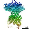


 PDBj
PDBj


 Assembly
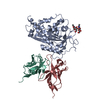
 Homo sapiens (human) / References: UniProt: A0A3Q3ANV4
 Sample preparation
 Electron microscopy imaging
 FIELD EMISSION GUN / Accelerating voltage: 300 kV / Illumination mode: FLOOD BEAM
 Processing