 Movie
Movie Controller


+ Open data

- Basic information
| Entry | Database: PDB / ID: 7lde | ||||||
|---|---|---|---|---|---|---|---|
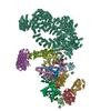
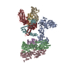
| Title | native AMPA receptor | ||||||
 Components Components |
| ||||||
 Keywords Keywords | MEMBRANE PROTEIN / neurotransmitter / hippocampus / ion-channel / glycosylation | ||||||
| Function / homology |  Function and homology information Function and homology informationPhase 0 - rapid depolarisation / Phase 2 - plateau phase / Activation of AMPA receptors / negative regulation of receptor localization to synapse / negative regulation of anterograde synaptic vesicle transport / Synaptic adhesion-like molecules / Cargo concentration in the ER / Unblocking of NMDA receptors, glutamate binding and activation / COPII-mediated vesicle transport / LGI-ADAM interactions ...Phase 0 - rapid depolarisation / Phase 2 - plateau phase / Activation of AMPA receptors / negative regulation of receptor localization to synapse / negative regulation of anterograde synaptic vesicle transport / Synaptic adhesion-like molecules / Cargo concentration in the ER / Unblocking of NMDA receptors, glutamate binding and activation / COPII-mediated vesicle transport / LGI-ADAM interactions / cellular response to amine stimulus / axonal spine / Trafficking of GluR2-containing AMPA receptors / Trafficking of AMPA receptors / positive regulation of membrane potential / neurotransmitter receptor activity involved in regulation of postsynaptic cytosolic calcium ion concentration / localization within membrane / cellular response to ammonium ion / neurotransmitter receptor transport, postsynaptic endosome to lysosome / L-type voltage-gated calcium channel complex / myosin V binding / neuron spine / cellular response to dsRNA / regulation of AMPA receptor activity / protein phosphatase 2B binding / neurotransmitter receptor internalization / response to arsenic-containing substance / postsynaptic neurotransmitter receptor diffusion trapping / nervous system process / dendritic spine membrane / long-term synaptic depression / cellular response to peptide hormone stimulus / beta-2 adrenergic receptor binding / protein kinase A binding / spinal cord development / neuronal cell body membrane / perisynaptic space / AMPA glutamate receptor activity / transmission of nerve impulse / channel regulator activity / response to lithium ion / immunoglobulin binding / regulation of postsynaptic membrane neurotransmitter receptor levels / AMPA glutamate receptor complex / excitatory synapse / adenylate cyclase binding / ionotropic glutamate receptor complex / regulation of postsynaptic membrane potential / postsynaptic density, intracellular component / asymmetric synapse / neuronal action potential / calcium channel regulator activity / regulation of receptor recycling / G-protein alpha-subunit binding / voltage-gated calcium channel activity / glutamate receptor binding / positive regulation of synaptic transmission / response to electrical stimulus / long-term memory / glutamate-gated receptor activity / response to fungicide / glutamate-gated calcium ion channel activity / presynaptic active zone membrane / vesicle-mediated transport / ionotropic glutamate receptor binding / somatodendritic compartment / dendrite membrane / synapse assembly / cellular response to brain-derived neurotrophic factor stimulus / ligand-gated monoatomic ion channel activity involved in regulation of presynaptic membrane potential / positive regulation of synaptic transmission, glutamatergic / regulation of membrane potential / dendritic shaft / response to cocaine / synaptic membrane / synaptic transmission, glutamatergic / long-term synaptic potentiation / cellular response to amino acid stimulus / transmitter-gated monoatomic ion channel activity involved in regulation of postsynaptic membrane potential / PDZ domain binding / postsynaptic density membrane / modulation of chemical synaptic transmission / neuromuscular junction / Schaffer collateral - CA1 synapse / regulation of synaptic plasticity / receptor internalization / cerebral cortex development / small GTPase binding / response to toxic substance / synaptic vesicle membrane / recycling endosome membrane / G-protein beta-subunit binding / cell-cell junction / synaptic vesicle / response to estradiol / presynapse / presynaptic membrane / amyloid-beta binding / early endosome membrane / cell body Similarity search - Function | ||||||
| Biological species |  | ||||||
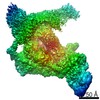
| Method | ELECTRON MICROSCOPY / single particle reconstruction / cryo EM / Resolution: 3.9 Å | ||||||
 Authors Authors | Yu, J. / Rao, P. / Gouaux, E. | ||||||
| Funding support |  United States, 1items United States, 1items
| ||||||
 Citation Citation | Journal: Nature / Year: 2021 Title: Hippocampal AMPA receptor assemblies and mechanism of allosteric inhibition. Authors: Jie Yu / Prashant Rao / Sarah Clark / Jaba Mitra / Taekjip Ha / Eric Gouaux / Abstract: AMPA-selective glutamate receptors mediate the transduction of signals between the neuronal circuits of the hippocampus. The trafficking, localization, kinetics and pharmacology of AMPA receptors are ...AMPA-selective glutamate receptors mediate the transduction of signals between the neuronal circuits of the hippocampus. The trafficking, localization, kinetics and pharmacology of AMPA receptors are tuned by an ensemble of auxiliary protein subunits, which are integral membrane proteins that associate with the receptor to yield bona fide receptor signalling complexes. Thus far, extensive studies of recombinant AMPA receptor-auxiliary subunit complexes using engineered protein constructs have not been able to faithfully elucidate the molecular architecture of hippocampal AMPA receptor complexes. Here we obtain mouse hippocampal, calcium-impermeable AMPA receptor complexes using immunoaffinity purification and use single-molecule fluorescence and cryo-electron microscopy experiments to elucidate three major AMPA receptor-auxiliary subunit complexes. The GluA1-GluA2, GluA1-GluA2-GluA3 and GluA2-GluA3 receptors are the predominant assemblies, with the auxiliary subunits TARP-γ8 and CNIH2-SynDIG4 non-stochastically positioned at the B'/D' and A'/C' positions, respectively. We further demonstrate how the receptor-TARP-γ8 stoichiometry explains the mechanism of and submaximal inhibition by a clinically relevant, brain-region-specific allosteric inhibitor. | ||||||
| History |
|
- Structure visualization
| Movie |
Movie viewer |
|---|---|
| Structure viewer | Molecule: MolmilJmol/JSmol |
- Downloads & links
-Download
| PDBx/mmCIF format | 7lde.cif.gz | 840.7 KB | Display | PDBx/mmCIF format |
|---|---|---|---|---|
| PDB format | pdb7lde.ent.gz | 647.3 KB | Display | PDB format |
| PDBx/mmJSON format | 7lde.json.gz | Tree view | PDBx/mmJSON format | |
| Others |  Other downloads Other downloads |
-Validation report
| Summary document | 7lde_validation.pdf.gz | 1.7 MB | Display | wwPDB validaton report |
|---|---|---|---|---|
| Full document | 7lde_full_validation.pdf.gz | 1.7 MB | Display | |
| Data in XML | 7lde_validation.xml.gz | 120.6 KB | Display | |
| Data in CIF | 7lde_validation.cif.gz | 185.2 KB | Display | |
| Arichive directory | https://data.pdbj.org/pub/pdb/validation_reports/ld/7ldeftp://data.pdbj.org/pub/pdb/validation_reports/ld/7lde | HTTPS FTP |
-Related structure data
| Related structure data |  23284MC  7lddC  7lepC M: map data used to model this data C: citing same article ( |
|---|---|
| Similar structure data |


-Links
 PDBj
PDBj


- Assembly
| Deposited unit | 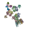
|
|---|---|
| 1 |
|
-Components
-Protein , 4 types, 8 molecules ACBDEFGH
| #1: Protein | Mass: 101678.969 Da / Num. of mol.: 2 / Source method: isolated from a natural source / Source: (natural) #2: Protein | Mass: 98899.883 Da / Num. of mol.: 2 / Source method: isolated from a natural source / Source: (natural) #3: Protein | Mass: 18948.420 Da / Num. of mol.: 2 / Source method: isolated from a natural source / Source: (natural) #4: Protein | Mass: 43502.938 Da / Num. of mol.: 2 / Source method: isolated from a natural source / Source: (natural) |
|---|
-Antibody , 3 types, 6 molecules ILJMKN
| #5: Antibody | Mass: 27511.527 Da / Num. of mol.: 2 Source method: isolated from a genetically manipulated source Source: (gene. exp.)  #6: Antibody | Mass: 25111.660 Da / Num. of mol.: 2 Source method: isolated from a genetically manipulated source Source: (gene. exp.) Production host:  Spodoptera aff. frugiperda 1 BOLD-2017 (butterflies/moths) Spodoptera aff. frugiperda 1 BOLD-2017 (butterflies/moths)#7: Antibody | Mass: 27975.439 Da / Num. of mol.: 2 Source method: isolated from a genetically manipulated source Source: (gene. exp.) Production host: Spodoptera aff. frugiperda 1 BOLD-2017 (butterflies/moths) |
|---|
-Sugars , 2 types, 12 molecules 
| #8: Polysaccharide | 2-acetamido-2-deoxy-beta-D-glucopyranose-(1-4)-2-acetamido-2-deoxy-beta-D-glucopyranose #14: Sugar | ChemComp-NAG /  Type: D-saccharide, beta linking / Mass: 221.208 Da / Num. of mol.: 7 / Source method: isolated from a natural source / Formula: C8H15NO6 Type: D-saccharide, beta linking / Mass: 221.208 Da / Num. of mol.: 7 / Source method: isolated from a natural source / Formula: C8H15NO6 |
|---|
-Non-polymers , 9 types, 40 molecules 


| #9: Chemical | ChemComp-ZK1 / {[  Mass: 409.254 Da / Num. of mol.: 4 / Source method: obtained synthetically / Formula: C14H15F3N3O6P / Comment: antagonist, medication*YM Mass: 409.254 Da / Num. of mol.: 4 / Source method: obtained synthetically / Formula: C14H15F3N3O6P / Comment: antagonist, medication*YM#10: Chemical |  Mass: 226.441 Da / Num. of mol.: 2 / Source method: obtained synthetically / Formula: C16H34 Mass: 226.441 Da / Num. of mol.: 2 / Source method: obtained synthetically / Formula: C16H34#11: Chemical | ChemComp-OCT /  Mass: 114.229 Da / Num. of mol.: 6 / Source method: obtained synthetically / Formula: C8H18 Mass: 114.229 Da / Num. of mol.: 6 / Source method: obtained synthetically / Formula: C8H18#12: Chemical | ChemComp-HP6 /  Mass: 100.202 Da / Num. of mol.: 4 / Source method: obtained synthetically / Formula: C7H16 Mass: 100.202 Da / Num. of mol.: 4 / Source method: obtained synthetically / Formula: C7H16#13: Chemical | ChemComp-D10 /  Mass: 142.282 Da / Num. of mol.: 6 / Source method: obtained synthetically / Formula: C10H22 Mass: 142.282 Da / Num. of mol.: 6 / Source method: obtained synthetically / Formula: C10H22#15: Chemical | ChemComp-D12 /  Mass: 170.335 Da / Num. of mol.: 6 / Source method: obtained synthetically / Formula: C12H26 Mass: 170.335 Da / Num. of mol.: 6 / Source method: obtained synthetically / Formula: C12H26#16: Chemical | ChemComp-C14 /  Mass: 198.388 Da / Num. of mol.: 6 / Source method: obtained synthetically / Formula: C14H30 Mass: 198.388 Da / Num. of mol.: 6 / Source method: obtained synthetically / Formula: C14H30#17: Chemical | ChemComp-DD9 /  Mass: 128.255 Da / Num. of mol.: 4 / Source method: obtained synthetically / Formula: C9H20 Mass: 128.255 Da / Num. of mol.: 4 / Source method: obtained synthetically / Formula: C9H20#18: Chemical |  Mass: 328.674 Da / Num. of mol.: 2 / Source method: obtained synthetically / Formula: C14H8ClF3N2O2 / Feature type: SUBJECT OF INVESTIGATION Mass: 328.674 Da / Num. of mol.: 2 / Source method: obtained synthetically / Formula: C14H8ClF3N2O2 / Feature type: SUBJECT OF INVESTIGATION |
|---|
-Details
| Has ligand of interest | Y |
|---|---|
| Has protein modification | Y |
-Experimental details
-Experiment
| Experiment | Method: ELECTRON MICROSCOPY |
|---|---|
| EM experiment | Aggregation state: PARTICLE / 3D reconstruction method: single particle reconstruction |
- Sample preparation
| Component |
| ||||||||||||||||||||||||||||||
|---|---|---|---|---|---|---|---|---|---|---|---|---|---|---|---|---|---|---|---|---|---|---|---|---|---|---|---|---|---|---|---|
| Source (natural) |
| ||||||||||||||||||||||||||||||
| Source (recombinant) |
| ||||||||||||||||||||||||||||||
| Buffer solution | pH: 8 | ||||||||||||||||||||||||||||||
| Specimen | Embedding applied: NO / Shadowing applied: NO / Staining applied: NO / Vitrification applied: YES | ||||||||||||||||||||||||||||||
| Vitrification | Cryogen name: ETHANE |
- Electron microscopy imaging
| Experimental equipment |  Model: Titan Krios / Image courtesy: FEI Company |
|---|---|
| Microscopy | Model: FEI TITAN KRIOS |
| Electron gun | Electron source:  FIELD EMISSION GUN / Accelerating voltage: 300 kV / Illumination mode: FLOOD BEAM FIELD EMISSION GUN / Accelerating voltage: 300 kV / Illumination mode: FLOOD BEAM |
| Electron lens | Mode: BRIGHT FIELD |
| Image recording | Electron dose: 50 e/Å2 / Film or detector model: GATAN K3 (6k x 4k) |
- Processing
| Software | Name: PHENIX / Version: 1.17_3644: / Classification: refinement | ||||||||||||||||||||||||
|---|---|---|---|---|---|---|---|---|---|---|---|---|---|---|---|---|---|---|---|---|---|---|---|---|---|
| CTF correction | Type: PHASE FLIPPING AND AMPLITUDE CORRECTION | ||||||||||||||||||||||||
| 3D reconstruction | Resolution: 3.9 Å / Resolution method: FSC 0.143 CUT-OFF / Num. of particles: 157000 / Symmetry type: POINT | ||||||||||||||||||||||||
| Refine LS restraints |
|
