 Movie
Movie Controller


+ Open data

- Basic information
| Entry | Database: PDB / ID: 7l0w | |||||||||||||||||||||||||||||||||
|---|---|---|---|---|---|---|---|---|---|---|---|---|---|---|---|---|---|---|---|---|---|---|---|---|---|---|---|---|---|---|---|---|---|---|
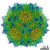
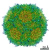
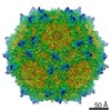
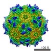
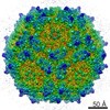
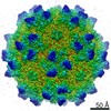
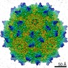
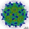
| Title | Human Bocavirus 1 (pH 5.5) | |||||||||||||||||||||||||||||||||
 Components Components | VP2 | |||||||||||||||||||||||||||||||||
 Keywords Keywords | VIRUS / Icosahedral Capsid / Human Bocavirus 1 / HBoV1 / Parvovirus | |||||||||||||||||||||||||||||||||
| Function / homology | Parvovirus coat protein VP2 / Parvovirus coat protein VP1/VP2 / Parvovirus coat protein VP1/VP2 / Capsid/spike protein, ssDNA virus / T=1 icosahedral viral capsid / structural molecule activity / VP2 Function and homology information Function and homology information | |||||||||||||||||||||||||||||||||
| Biological species | Primate bocaparvovirus 1 | |||||||||||||||||||||||||||||||||
| Method | ELECTRON MICROSCOPY / single particle reconstruction / cryo EM / Resolution: 2.74 Å | |||||||||||||||||||||||||||||||||
 Authors Authors | Luo, M. / Mietzsch, M. / Agbandje-McKenna, M. | |||||||||||||||||||||||||||||||||
| Funding support |  United States, 1items United States, 1items
| |||||||||||||||||||||||||||||||||
 Citation Citation | Journal: J Virol / Year: 2021 Title: pH-Induced Conformational Changes of Human Bocavirus Capsids. Authors: Mengxiao Luo / Mario Mietzsch / Paul Chipman / Kangkang Song / Chen Xu / John Spear / Duncan Sousa / Robert McKenna / Maria Söderlund-Venermo / Mavis Agbandje-McKenna /  Abstract: Human bocavirus 1 (HBoV1) and HBoV2-4 infect children and immunocompromised individuals, resulting in respiratory and gastrointestinal infections, respectively. Using cryo-electron microscopy and ...Human bocavirus 1 (HBoV1) and HBoV2-4 infect children and immunocompromised individuals, resulting in respiratory and gastrointestinal infections, respectively. Using cryo-electron microscopy and image reconstruction, the HBoV2 capsid structure was determined to 2.7 Å resolution at pH 7.4 and compared to the previously determined HBoV1, HBoV3, and HBoV4 structures. Consistent with previous findings, surface variable region (VR) III of the capsid protein VP3, proposed as a host tissue-tropism determinant, was structurally similar among the gastrointestinal strains HBoV2-4, but differed from HBoV1 with its tropism for the respiratory tract. Towards understanding the entry and trafficking properties of these viruses, HBoV1 and HBoV2 were further analyzed as species representatives of the two HBoV tropisms. Their cell surface glycan-binding characteristics were analyzed, and capsid structures determined to 2.5-2.7 Å resolution at pH 5.5 and 2.6, conditions normally encountered during infection. The data showed that glycans with terminal sialic acid, galactose, GlcNAc or heparan sulfate moieties do not facilitate HBoV1 or HBoV2 cellular attachment. With respect to trafficking, conformational changes common to both viruses were observed at low pH conditions localized to the VP N-terminus under the 5-fold channel, in the surface loops VR-I and VR-V and specific side-chain residues such as cysteines and histidines. The 5-fold conformational movements provide insight into the potential mechanism of VP N-terminal dynamics during HBoV infection and side-chain modifications highlight pH-sensitive regions of the capsid. Human bocaviruses (HBoVs) are associated with disease in humans. However, the lack of an animal model and a versatile cell culture system to study their life cycle limits the ability to develop specific treatments or vaccines. This study presents the structure of HBoV2, at 2.7 Å resolution, determined for comparison to the existing HBoV1, HBoV3, and HBoV4 structures, to enable the molecular characterization of strain and genus-specific capsid features contributing to tissue tropism and antigenicity. Furthermore, HBoV1 and HBoV2 structures determined under acidic conditions provide insight into capsid changes associated with endosomal and gastrointestinal acidification. Structural rearrangements of the capsid VP N-terminus, at the base of the 5-fold channel, demonstrate a disordering of a "basket" motif as pH decreases. These observations begin to unravel the molecular mechanism of HBoV infection and provide information for control strategies. | |||||||||||||||||||||||||||||||||
| History |
|
- Structure visualization
| Movie |
Movie viewer |
|---|---|
| Structure viewer | Molecule: MolmilJmol/JSmol |
- Downloads & links
-Download
| PDBx/mmCIF format | 7l0w.cif.gz | 5 MB | Display | PDBx/mmCIF format |
|---|---|---|---|---|
| PDB format | pdb7l0w.ent.gz | Display | PDB format | |
| PDBx/mmJSON format | 7l0w.json.gz | Tree view | PDBx/mmJSON format | |
| Others |  Other downloads Other downloads |
-Validation report
| Arichive directory | https://data.pdbj.org/pub/pdb/validation_reports/l0/7l0wftp://data.pdbj.org/pub/pdb/validation_reports/l0/7l0w | HTTPS FTP |
|---|
-Related structure data
| Related structure data |  23106MC  7l0uC  7l0vC  7l0xC  7l0yC M: map data used to model this data C: citing same article ( |
|---|---|
| Similar structure data |


-Links
 PDBj
PDBj


- Assembly
| Deposited unit | 
|
|---|---|
| 1 |
|
-Components
| #1: Protein | Mass: 57450.082 Da / Num. of mol.: 60 / Fragment: UNP residues 33-542 Source method: isolated from a genetically manipulated source Source: (gene. exp.)  Primate bocaparvovirus 1 (strain Human bocavirus 1 type 1) Primate bocaparvovirus 1 (strain Human bocavirus 1 type 1)Strain: Human bocavirus 1 type 1 / Gene: vp2, VP2 / Cell line (production host): Sf9 / Production host:   Spodoptera frugiperda (fall armyworm) / References: UniProt: H9C5X6 Spodoptera frugiperda (fall armyworm) / References: UniProt: H9C5X6Has protein modification | N | |
|---|
-Experimental details
-Experiment
| Experiment | Method: ELECTRON MICROSCOPY |
|---|---|
| EM experiment | Aggregation state: PARTICLE / 3D reconstruction method: single particle reconstruction |
- Sample preparation
| Component | Name: Human bocavirus 1 / Type: VIRUS / Entity ID: all / Source: RECOMBINANT |
|---|---|
| Source (natural) | Organism: Human bocavirus 1 |
| Source (recombinant) | Organism: Spodoptera frugiperda (fall armyworm) / Cell: Sf9 |
| Details of virus | Empty: NO / Enveloped: NO / Isolate: OTHER / Type: VIRION |
| Buffer solution | pH: 5.5 |
| Specimen | Embedding applied: NO / Shadowing applied: NO / Staining applied: NO / Vitrification applied: YES |
| Vitrification | Cryogen name: ETHANE |
- Electron microscopy imaging
| Experimental equipment |  Model: Titan Krios / Image courtesy: FEI Company |
|---|---|
| Microscopy | Model: FEI TITAN KRIOS |
| Electron gun | Electron source:  FIELD EMISSION GUN / Accelerating voltage: 300 kV / Illumination mode: FLOOD BEAM FIELD EMISSION GUN / Accelerating voltage: 300 kV / Illumination mode: FLOOD BEAM |
| Electron lens | Mode: BRIGHT FIELD / Cs: 2.7 mm |
| Image recording | Electron dose: 64 e/Å2 / Film or detector model: GATAN K2 SUMMIT (4k x 4k) |
- Processing
| Software | Name: PHENIX / Version: 1.10-2155_2155: / Classification: refinement | ||||||||||||||||||||||||
|---|---|---|---|---|---|---|---|---|---|---|---|---|---|---|---|---|---|---|---|---|---|---|---|---|---|
| EM software |
| ||||||||||||||||||||||||
| CTF correction | Type: PHASE FLIPPING AND AMPLITUDE CORRECTION | ||||||||||||||||||||||||
| 3D reconstruction | Resolution: 2.74 Å / Resolution method: FSC 0.143 CUT-OFF / Num. of particles: 66471 / Symmetry type: POINT | ||||||||||||||||||||||||
| Refine LS restraints |
|
