Movie
Movie Controller


+ Open data

- Basic information
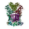
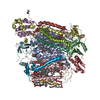
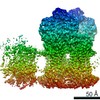
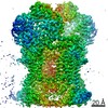
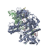
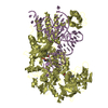
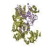
| Entry | Database: PDB / ID: 7jro | ||||||
|---|---|---|---|---|---|---|---|
| Title | Plant Mitochondrial complex IV from Vigna radiata | ||||||
 Components Components |
| ||||||
 Keywords Keywords | OXIDOREDUCTASE / mitochondria / respiration / bioenergetics / plants | ||||||
| Function / homology |  Function and homology information Function and homology informationrespiratory chain complex IV / mitochondrial envelope / transmembrane transporter complex / mitochondrial electron transport, cytochrome c to oxygen / cytochrome-c oxidase activity / catalytic complex / ATP synthesis coupled electron transport / enzyme regulator activity / oxidoreductase activity / mitochondrial inner membrane ...respiratory chain complex IV / mitochondrial envelope / transmembrane transporter complex / mitochondrial electron transport, cytochrome c to oxygen / cytochrome-c oxidase activity / catalytic complex / ATP synthesis coupled electron transport / enzyme regulator activity / oxidoreductase activity / mitochondrial inner membrane / copper ion binding / mitochondrion / metal ion binding Similarity search - Function | ||||||
| Biological species |  Vigna radiata (mung bean)Vigna radiata var. radiata (mung bean) Vigna radiata (mung bean)Vigna radiata var. radiata (mung bean) | ||||||
| Method | ELECTRON MICROSCOPY / single particle reconstruction / cryo EM / Resolution: 3.8 Å | ||||||
 Authors Authors | Maldonado, M. / Letts, J.A. | ||||||
 Citation Citation | Journal: Elife / Year: 2021 Title: Atomic structures of respiratory complex III, complex IV, and supercomplex III-IV from vascular plants. Authors: Maria Maldonado / Fei Guo / James A Letts /  Abstract: Mitochondrial complex III (CIII) and complex IV (CIV), which can associate into a higher-order supercomplex (SC III+IV), play key roles in respiration. However, structures of these plant complexes ...Mitochondrial complex III (CIII) and complex IV (CIV), which can associate into a higher-order supercomplex (SC III+IV), play key roles in respiration. However, structures of these plant complexes remain unknown. We present atomic models of CIII, CIV, and SC III+IV from determined by single-particle cryoEM. The structures reveal plant-specific differences in the MPP domain of CIII and define the subunit composition of CIV. Conformational heterogeneity analysis of CIII revealed long-range, coordinated movements across the complex, as well as the motion of CIII's iron-sulfur head domain. The CIV structure suggests that, in plants, proton translocation does not occur via the H channel. The supercomplex interface differs significantly from that in yeast and bacteria in its interacting subunits, angle of approach and limited interactions in the mitochondrial matrix. These structures challenge long-standing assumptions about the plant complexes and generate new mechanistic hypotheses. | ||||||
| History |
|
- Structure visualization
| Movie |
Movie viewer |
|---|---|
| Structure viewer | Molecule: MolmilJmol/JSmol |
- Downloads & links
-Download
| PDBx/mmCIF format | 7jro.cif.gz | 308.1 KB | Display | PDBx/mmCIF format |
|---|---|---|---|---|
| PDB format | pdb7jro.ent.gz | 242.8 KB | Display | PDB format |
| PDBx/mmJSON format | 7jro.json.gz | Tree view | PDBx/mmJSON format | |
| Others |  Other downloads Other downloads |
-Validation report
| Arichive directory | https://data.pdbj.org/pub/pdb/validation_reports/jr/7jroftp://data.pdbj.org/pub/pdb/validation_reports/jr/7jro | HTTPS FTP |
|---|
-Related structure data
| Related structure data | 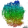 22447MC  7jrgC 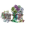 7jrpC C: citing same article ( M: map data used to model this data |
|---|---|
| Similar structure data | |
| EM raw data | EMPIAR-10586 (Title: Cryo electron micrographs of digitonin-solubilized, amphipol-stabilized, sucrose-gradient-purified V. radiata mitochondrial membranes - mixed fraction containing CI*, CIII2 and SC III2+IV Data size: 6.7 TB Data #1: Raw movies of mixed sample used for determination of mung bean respiratory CI*, CIII2, CIV and SC III2+IV [micrographs - multiframe]) |


-Links
 PDBj
PDBj


- Assembly
| Deposited unit | 
|
|---|---|
| 1 |
|
-Components
-Protein , 5 types, 5 molecules acdij
| #1: Protein | Mass: 57960.559 Da / Num. of mol.: 1 / Source method: isolated from a natural source / Source: (natural) Vigna radiata (mung bean) |
|---|---|
| #3: Protein | Mass: 29967.893 Da / Num. of mol.: 1 / Source method: isolated from a natural source / Source: (natural) Vigna radiata (mung bean) |
| #4: Protein | Mass: 8811.020 Da / Num. of mol.: 1 / Source method: isolated from a natural source / Source: (natural) Vigna radiata var. radiata (mung bean) / References: UniProt: A0A1S3TNX9 |
| #9: Protein | Mass: 7622.931 Da / Num. of mol.: 1 / Source method: isolated from a natural source / Source: (natural) Vigna radiata var. radiata (mung bean) / References: UniProt: A0A1S3TUX2 |
| #10: Protein | Mass: 10505.158 Da / Num. of mol.: 1 / Source method: isolated from a natural source / Source: (natural) Vigna radiata var. radiata (mung bean) / References: UniProt: A0A1S3V7C2 |
-Cytochrome c oxidase subunit ... , 5 types, 5 molecules befgh
| #2: Protein | Mass: 28391.602 Da / Num. of mol.: 1 / Source method: isolated from a natural source / Source: (natural) Vigna radiata var. radiata (mung bean) / References: UniProt: A0A1S3TZB2 |
|---|---|
| #5: Protein | Mass: 16694.705 Da / Num. of mol.: 1 / Source method: isolated from a natural source / Source: (natural) Vigna radiata var. radiata (mung bean) / References: UniProt: A0A1S3U1E4 |
| #6: Protein | Mass: 11225.671 Da / Num. of mol.: 1 / Source method: isolated from a natural source / Source: (natural) Vigna radiata var. radiata (mung bean) / References: UniProt: A0A1S3UMA0 |
| #7: Protein | Mass: 20018.635 Da / Num. of mol.: 1 / Source method: isolated from a natural source / Source: (natural) Vigna radiata var. radiata (mung bean) / References: UniProt: A0A1S3VV60 |
| #8: Protein | Mass: 7120.326 Da / Num. of mol.: 1 / Source method: isolated from a natural source / Source: (natural) Vigna radiata var. radiata (mung bean) / References: UniProt: A0A1S3V319 |
-Non-polymers , 9 types, 27 molecules 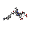


| #11: Chemical |  Mass: 852.837 Da / Num. of mol.: 2 / Source method: obtained synthetically / Formula: C49H56FeN4O6 Mass: 852.837 Da / Num. of mol.: 2 / Source method: obtained synthetically / Formula: C49H56FeN4O6#12: Chemical | ChemComp-CU / |  Mass: 63.546 Da / Num. of mol.: 1 / Source method: obtained synthetically / Formula: Cu Mass: 63.546 Da / Num. of mol.: 1 / Source method: obtained synthetically / Formula: Cu#13: Chemical | ChemComp-MG / |  Mass: 24.305 Da / Num. of mol.: 1 / Source method: obtained synthetically / Formula: Mg Mass: 24.305 Da / Num. of mol.: 1 / Source method: obtained synthetically / Formula: Mg#14: Chemical |  Mass: 790.145 Da / Num. of mol.: 2 / Source method: obtained synthetically / Formula: C44H88NO8P / Comment: phospholipid*YM Mass: 790.145 Da / Num. of mol.: 2 / Source method: obtained synthetically / Formula: C44H88NO8P / Comment: phospholipid*YM#15: Chemical | ChemComp-3PE /  Mass: 748.065 Da / Num. of mol.: 17 / Source method: obtained synthetically / Formula: C41H82NO8P / Comment: phospholipid*YM Mass: 748.065 Da / Num. of mol.: 17 / Source method: obtained synthetically / Formula: C41H82NO8P / Comment: phospholipid*YM#16: Chemical | ChemComp-CDL / |  Mass: 1464.043 Da / Num. of mol.: 1 / Source method: obtained synthetically / Formula: C81H156O17P2 / Comment: phospholipid*YM Mass: 1464.043 Da / Num. of mol.: 1 / Source method: obtained synthetically / Formula: C81H156O17P2 / Comment: phospholipid*YM#17: Chemical | ChemComp-CUA / |  Mass: 127.092 Da / Num. of mol.: 1 / Source method: obtained synthetically / Formula: Cu2 Mass: 127.092 Da / Num. of mol.: 1 / Source method: obtained synthetically / Formula: Cu2#18: Chemical | ChemComp-ZN / |  Mass: 65.409 Da / Num. of mol.: 1 / Source method: obtained synthetically / Formula: Zn Mass: 65.409 Da / Num. of mol.: 1 / Source method: obtained synthetically / Formula: Zn#19: Chemical | ChemComp-LYS / |  Type: L-peptide linking / Mass: 147.195 Da / Num. of mol.: 1 / Source method: obtained synthetically / Formula: C6H15N2O2 Type: L-peptide linking / Mass: 147.195 Da / Num. of mol.: 1 / Source method: obtained synthetically / Formula: C6H15N2O2 |
|---|
-Details
| Has ligand of interest | N |
|---|---|
| Has protein modification | Y |
-Experimental details
-Experiment
| Experiment | Method: ELECTRON MICROSCOPY |
|---|---|
| EM experiment | Aggregation state: PARTICLE / 3D reconstruction method: single particle reconstruction |
- Sample preparation
| Component | Name: Mitochondrial Respiratory Complex IV / Type: COMPLEX / Entity ID: #1-#10 / Source: NATURAL | |||||||||||||||||||||||||
|---|---|---|---|---|---|---|---|---|---|---|---|---|---|---|---|---|---|---|---|---|---|---|---|---|---|---|
| Molecular weight | Value: 0.485 MDa / Experimental value: NO | |||||||||||||||||||||||||
| Source (natural) | Organism: Vigna radiata (mung bean) / Organelle: mitochondria / Tissue: hypocotyl | |||||||||||||||||||||||||
| Buffer solution | pH: 7.8 | |||||||||||||||||||||||||
| Buffer component |
| |||||||||||||||||||||||||
| Specimen | Conc.: 6 mg/ml / Embedding applied: NO / Shadowing applied: NO / Staining applied: NO / Vitrification applied: YES Details: This sample was monodisperse on size exclusion chromatography but was a mixture of different mitochondrial respiratory complexes | |||||||||||||||||||||||||
| Specimen support | Grid material: COPPER / Grid mesh size: 300 divisions/in. / Grid type: Quantifoil R1.2/1.3 | |||||||||||||||||||||||||
| Vitrification | Instrument: FEI VITROBOT MARK III / Cryogen name: ETHANE / Humidity: 100 % / Chamber temperature: 288 K |
- Electron microscopy imaging
| Experimental equipment |  Model: Titan Krios / Image courtesy: FEI Company |
|---|---|
| Microscopy | Model: FEI TITAN KRIOS |
| Electron gun | Electron source:  FIELD EMISSION GUN / Accelerating voltage: 300 kV / Illumination mode: FLOOD BEAM FIELD EMISSION GUN / Accelerating voltage: 300 kV / Illumination mode: FLOOD BEAM |
| Electron lens | Mode: BRIGHT FIELD / Calibrated magnification: 60010 X / Calibrated defocus min: 1500 nm / Calibrated defocus max: 3000 nm / Alignment procedure: COMA FREE |
| Specimen holder | Cryogen: NITROGEN / Specimen holder model: FEI TITAN KRIOS AUTOGRID HOLDER |
| Image recording | Average exposure time: 3 sec. / Electron dose: 86.4 e/Å2 / Film or detector model: GATAN K3 (6k x 4k) / Num. of grids imaged: 1 / Num. of real images: 9816 |
| Image scans | Width: 5760 / Height: 4092 |
- Processing
| EM software |
| ||||||||||||||||||||||||||||||||||||
|---|---|---|---|---|---|---|---|---|---|---|---|---|---|---|---|---|---|---|---|---|---|---|---|---|---|---|---|---|---|---|---|---|---|---|---|---|---|
| CTF correction | Type: PHASE FLIPPING AND AMPLITUDE CORRECTION | ||||||||||||||||||||||||||||||||||||
| Particle selection | Num. of particles selected: 150000000 | ||||||||||||||||||||||||||||||||||||
| Symmetry | Point symmetry: C1 (asymmetric) | ||||||||||||||||||||||||||||||||||||
| 3D reconstruction | Resolution: 3.8 Å / Resolution method: FSC 0.143 CUT-OFF / Num. of particles: 29348 / Algorithm: FOURIER SPACE / Symmetry type: POINT | ||||||||||||||||||||||||||||||||||||
| Atomic model building | B value: 67 / Protocol: AB INITIO MODEL / Space: REAL / Target criteria: correlation coefficient | ||||||||||||||||||||||||||||||||||||
| Atomic model building |
|