ムービー
ムービー コントローラー


+ データを開く

- 基本情報
基本情報
| 登録情報 | データベース: PDB / ID: 7bti | ||||||||||||||||||||||||||||||||||||
|---|---|---|---|---|---|---|---|---|---|---|---|---|---|---|---|---|---|---|---|---|---|---|---|---|---|---|---|---|---|---|---|---|---|---|---|---|---|
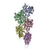
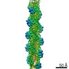
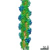
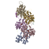
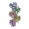
| タイトル | Phalloidin bound F-actin complex | ||||||||||||||||||||||||||||||||||||
 要素 要素 |
| ||||||||||||||||||||||||||||||||||||
 キーワード キーワード | CONTRACTILE PROTEIN/PROTEIN BINDING / F-actin / ADP-F-actin / CONTRACTILE PROTEIN / CONTRACTILE PROTEIN-PROTEIN BINDING complex | ||||||||||||||||||||||||||||||||||||
| 機能・相同性 |  機能・相同性情報 機能・相同性情報Striated Muscle Contraction / striated muscle thin filament / skeletal muscle thin filament assembly / skeletal muscle fiber development / stress fiber / actin filament / 加水分解酵素; 酸無水物に作用; 酸無水物に作用・細胞または細胞小器官の運動に関与 / actin cytoskeleton / toxin activity / hydrolase activity / ATP binding 類似検索 - 分子機能 | ||||||||||||||||||||||||||||||||||||
| 生物種 |   Amanita phalloides (タマゴテングタケ) Amanita phalloides (タマゴテングタケ) | ||||||||||||||||||||||||||||||||||||
| 手法 | 電子顕微鏡法 / らせん対称体再構成法 / クライオ電子顕微鏡法 / 解像度: 3.6 Å | ||||||||||||||||||||||||||||||||||||
 データ登録者 データ登録者 | Kumari, A. / Ragunath, V.K. / Sirajuddin, M. | ||||||||||||||||||||||||||||||||||||
| 資金援助 |  インド, 2件 インド, 2件
| ||||||||||||||||||||||||||||||||||||
 引用 引用 | ジャーナル: EMBO J / 年: 2020 タイトル: Structural insights into actin filament recognition by commonly used cellular actin markers. 著者: Archana Kumari / Shubham Kesarwani / Manjunath G Javoor / Kutti R Vinothkumar / Minhajuddin Sirajuddin / 要旨: Cellular studies of filamentous actin (F-actin) processes commonly utilize fluorescent versions of toxins, peptides, and proteins that bind actin. While the choice of these markers has been largely ...Cellular studies of filamentous actin (F-actin) processes commonly utilize fluorescent versions of toxins, peptides, and proteins that bind actin. While the choice of these markers has been largely based on availability and ease, there is a severe dearth of structural data for an informed judgment in employing suitable F-actin markers for a particular requirement. Here, we describe the electron cryomicroscopy structures of phalloidin, lifeAct, and utrophin bound to F-actin, providing a comprehensive high-resolution structural comparison of widely used actin markers and their influence towards F-actin. Our results show that phalloidin binding does not induce specific conformational change and lifeAct specifically recognizes closed D-loop conformation, i.e., ADP-Pi or ADP states of F-actin. The structural models aided designing of minimal utrophin and a shorter lifeAct, which can be utilized as F-actin marker. Together, our study provides a structural perspective, where the binding sites of utrophin and lifeAct overlap with majority of actin-binding proteins and thus offering an invaluable resource for researchers in choosing appropriate actin markers and generating new marker variants. | ||||||||||||||||||||||||||||||||||||
| 履歴 |
|
- 構造の表示
構造の表示
| ムービー |
ムービービューア |
|---|---|
| 構造ビューア | 分子: MolmilJmol/JSmol |
- ダウンロードとリンク
-ダウンロード
| PDBx/mmCIF形式 | 7bti.cif.gz | 335 KB | 表示 | PDBx/mmCIF形式 |
|---|---|---|---|---|
| PDB形式 | pdb7bti.ent.gz | 266.8 KB | 表示 | PDB形式 |
| PDBx/mmJSON形式 | 7bti.json.gz | ツリー表示 | PDBx/mmJSON形式 | |
| その他 |  その他のダウンロード その他のダウンロード |
-検証レポート
| 文書・要旨 | 7bti_validation.pdf.gz | 681.2 KB | 表示 | wwPDB検証レポート |
|---|---|---|---|---|
| 文書・詳細版 | 7bti_full_validation.pdf.gz | 713.1 KB | 表示 | |
| XML形式データ | 7bti_validation.xml.gz | 39.8 KB | 表示 | |
| CIF形式データ | 7bti_validation.cif.gz | 57 KB | 表示 | |
| アーカイブディレクトリ | https://data.pdbj.org/pub/pdb/validation_reports/bt/7btiftp://data.pdbj.org/pub/pdb/validation_reports/bt/7bti | HTTPS FTP |
-関連構造データ


-リンク
 PDBj
PDBj


- 集合体
集合体
| 登録構造単位 | 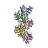
|
|---|---|
| 1 |
|
-要素
| #1: タンパク質 | 分子量: 42096.953 Da / 分子数: 5 / 由来タイプ: 天然 / 由来: (天然) #2: タンパク質・ペプチド | 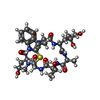  タイプ: Cyclic peptide / クラス: 毒素 / 分子量: 808.899 Da / 分子数: 3 / 由来タイプ: 合成 タイプ: Cyclic peptide / クラス: 毒素 / 分子量: 808.899 Da / 分子数: 3 / 由来タイプ: 合成由来: (合成) Amanita phalloides (タマゴテングタケ)参照: Phalloidin, UniProt: P0CU64*PLUS #3: 化合物 | ChemComp-MG /   分子量: 24.305 Da / 分子数: 5 / 由来タイプ: 合成 / 式: Mg / タイプ: SUBJECT OF INVESTIGATION 分子量: 24.305 Da / 分子数: 5 / 由来タイプ: 合成 / 式: Mg / タイプ: SUBJECT OF INVESTIGATION#4: 化合物 | ChemComp-ADP /   分子量: 427.201 Da / 分子数: 5 / 由来タイプ: 合成 / 式: C10H15N5O10P2 / タイプ: SUBJECT OF INVESTIGATION / コメント: ADP, エネルギー貯蔵分子*YM 分子量: 427.201 Da / 分子数: 5 / 由来タイプ: 合成 / 式: C10H15N5O10P2 / タイプ: SUBJECT OF INVESTIGATION / コメント: ADP, エネルギー貯蔵分子*YM研究の焦点であるリガンドがあるか | Y | Has protein modification | Y | |
|---|
-実験情報
-実験
| 実験 | 手法: 電子顕微鏡法 |
|---|---|
| EM実験 | 試料の集合状態: FILAMENT / 3次元再構成法: らせん対称体再構成法 |
- 試料調製
試料調製
| 構成要素 | 名称: Filamentous actin in ADP state / タイプ: COMPLEX / Entity ID: #1-#2 / 由来: NATURAL |
|---|---|
| 分子量 | 実験値: NO |
| 由来(天然) | 生物種: |
| 緩衝液 | pH: 7.5 詳細: 50mM KCl,1mM MgCl2,0.2mM EGTA, 10mM Imidazole buffer pH 7.5 |
| 試料 | 濃度: 0.0002 mg/ml / 包埋: NO / シャドウイング: NO / 染色: NO / 凍結: YES 詳細: 10 mole excess of lifeact was mixed with F-actin and used for sample preparation. |
| 試料支持 | グリッドの材料: GOLD / グリッドのサイズ: 300 divisions/in. / グリッドのタイプ: Quantifoil R1.2/1.3 |
| 急速凍結 | 装置: FEI VITROBOT MARK I / 凍結剤: ETHANE / 湿度: 95 % / 凍結前の試料温度: 293 K / 詳細: blot for 3.5 seconds |
- 電子顕微鏡撮影
| 実験機器 |  モデル: Titan Krios / 画像提供: FEI Company |
|---|---|
| 顕微鏡 | モデル: FEI TITAN KRIOS |
| 電子銃 | 電子線源:  FIELD EMISSION GUN / 加速電圧: 300 kV / 照射モード: OTHER FIELD EMISSION GUN / 加速電圧: 300 kV / 照射モード: OTHER |
| 電子レンズ | モード: OTHER / 倍率(補正後): 75000 X / Cs: 2.7 mm / C2レンズ絞り径: 50 µm / アライメント法: COMA FREE |
| 試料ホルダ | 凍結剤: NITROGEN 試料ホルダーモデル: FEI TITAN KRIOS AUTOGRID HOLDER 最高温度: 120 K / 最低温度: 100 K |
| 撮影 | 平均露光時間: 2 sec. / 電子線照射量: 49.2 e/Å2 / 検出モード: INTEGRATING フィルム・検出器のモデル: FEI FALCON III (4k x 4k) 撮影したグリッド数: 1 / 実像数: 529 / 詳細: 30 |
- 解析
解析
| ソフトウェア |
| ||||||||||||||||||||||||||||||||||||||||||||||||||
|---|---|---|---|---|---|---|---|---|---|---|---|---|---|---|---|---|---|---|---|---|---|---|---|---|---|---|---|---|---|---|---|---|---|---|---|---|---|---|---|---|---|---|---|---|---|---|---|---|---|---|---|
| EMソフトウェア |
| ||||||||||||||||||||||||||||||||||||||||||||||||||
| CTF補正 | 詳細: GCTF for CTF correction / タイプ: NONE | ||||||||||||||||||||||||||||||||||||||||||||||||||
| らせん対称 | 回転角度/サブユニット: -167 ° / 軸方向距離/サブユニット: 27.89 Å / らせん対称軸の対称性: C1 | ||||||||||||||||||||||||||||||||||||||||||||||||||
| 粒子像の選択 | 選択した粒子像数: 349839 | ||||||||||||||||||||||||||||||||||||||||||||||||||
| 3次元再構成 | 解像度: 3.6 Å / 解像度の算出法: FSC 0.143 CUT-OFF / 粒子像の数: 91245 / クラス平均像の数: 4 / 対称性のタイプ: HELICAL | ||||||||||||||||||||||||||||||||||||||||||||||||||
| 原子モデル構築 | B value: 179 / プロトコル: RIGID BODY FIT / 空間: REAL | ||||||||||||||||||||||||||||||||||||||||||||||||||
| 原子モデル構築 | PDB-ID: 5ONV PDB chain-ID: A / Accession code: 5ONV / Pdb chain residue range: 6-373 / Source name: PDB / タイプ: experimental model | ||||||||||||||||||||||||||||||||||||||||||||||||||
| 精密化 | 立体化学のターゲット値: GeoStd + Monomer Library + CDL v1.2 | ||||||||||||||||||||||||||||||||||||||||||||||||||
| 拘束条件 |
|