 Movie
Movie Controller


+ Open data

- Basic information
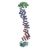
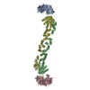
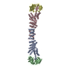
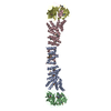
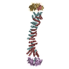
| Entry | Database: PDB / ID: 6zl0 | |||||||||
|---|---|---|---|---|---|---|---|---|---|---|
| Title | COPII on membranes, outer coat left-handed rod | |||||||||
 Components Components |
| |||||||||
 Keywords Keywords | PROTEIN TRANSPORT / SECRETION / TRAFFICKING | |||||||||
| Function / homology |  Function and homology information Function and homology informationpositive regulation of ER to Golgi vesicle-mediated transport / Seh1-associated complex / COPII-coated vesicle budding / nuclear pore localization / protein exit from endoplasmic reticulum / COPII-mediated vesicle transport / regulation of TORC1 signaling / nuclear pore outer ring / COPII-coated vesicle cargo loading / Regulation of Glucokinase by Glucokinase Regulatory Protein ...positive regulation of ER to Golgi vesicle-mediated transport / Seh1-associated complex / COPII-coated vesicle budding / nuclear pore localization / protein exit from endoplasmic reticulum / COPII-mediated vesicle transport / regulation of TORC1 signaling / nuclear pore outer ring / COPII-coated vesicle cargo loading / Regulation of Glucokinase by Glucokinase Regulatory Protein / : / positive regulation of protein exit from endoplasmic reticulum / Regulation of HSF1-mediated heat shock response / COPII vesicle coat / SUMOylation of SUMOylation proteins / mating projection tip / endoplasmic reticulum organization / SUMOylation of RNA binding proteins / SUMOylation of chromatin organization proteins / vacuolar membrane / nucleocytoplasmic transport / endoplasmic reticulum exit site / positive regulation of TOR signaling / nuclear pore / mRNA transport / ERAD pathway / positive regulation of TORC1 signaling / cell periphery / protein import into nucleus / nuclear envelope / protein transport / endoplasmic reticulum membrane / positive regulation of DNA-templated transcription / structural molecule activity / endoplasmic reticulum Similarity search - Function | |||||||||
| Biological species |  | |||||||||
| Method | ELECTRON MICROSCOPY / subtomogram averaging / cryo EM / Resolution: 40 Å | |||||||||
 Authors Authors | Zanetti, G. / Hutchings, J. | |||||||||
| Funding support |  United Kingdom, 2items United Kingdom, 2items
| |||||||||
 Citation Citation | Journal: Nat Commun / Year: 2021 Title: Structure of the complete, membrane-assembled COPII coat reveals a complex interaction network. Authors: Joshua Hutchings / Viktoriya G Stancheva / Nick R Brown / Alan C M Cheung / Elizabeth A Miller / Giulia Zanetti /  Abstract: COPII mediates Endoplasmic Reticulum to Golgi trafficking of thousands of cargoes. Five essential proteins assemble into a two-layer architecture, with the inner layer thought to regulate coat ...COPII mediates Endoplasmic Reticulum to Golgi trafficking of thousands of cargoes. Five essential proteins assemble into a two-layer architecture, with the inner layer thought to regulate coat assembly and cargo recruitment, and the outer coat forming cages assumed to scaffold membrane curvature. Here we visualise the complete, membrane-assembled COPII coat by cryo-electron tomography and subtomogram averaging, revealing the full network of interactions within and between coat layers. We demonstrate the physiological importance of these interactions using genetic and biochemical approaches. Mutagenesis reveals that the inner coat alone can provide membrane remodelling function, with organisational input from the outer coat. These functional roles for the inner and outer coats significantly move away from the current paradigm, which posits membrane curvature derives primarily from the outer coat. We suggest these interactions collectively contribute to coat organisation and membrane curvature, providing a structural framework to understand regulatory mechanisms of COPII trafficking and secretion. | |||||||||
| History |
|
- Structure visualization
| Movie |
Movie viewer |
|---|---|
| Structure viewer | Molecule: MolmilJmol/JSmol |
- Downloads & links
-Download
| PDBx/mmCIF format | 6zl0.cif.gz | 283.5 KB | Display | PDBx/mmCIF format |
|---|---|---|---|---|
| PDB format | pdb6zl0.ent.gz | 206.8 KB | Display | PDB format |
| PDBx/mmJSON format | 6zl0.json.gz | Tree view | PDBx/mmJSON format | |
| Others |  Other downloads Other downloads |
-Validation report
| Arichive directory | https://data.pdbj.org/pub/pdb/validation_reports/zl/6zl0ftp://data.pdbj.org/pub/pdb/validation_reports/zl/6zl0 | HTTPS FTP |
|---|
-Related structure data
| Related structure data |  11264MC 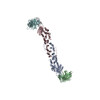 6zg5C  6zg6C  6zgaC M: map data used to model this data C: citing same article ( |
|---|---|
| Similar structure data |


-Links
 PDBj
PDBj

- Assembly
| Deposited unit | 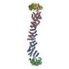
|
|---|---|
| 1 |
|
-Components
| #1: Protein | Mass: 138847.438 Da / Num. of mol.: 2 Source method: isolated from a genetically manipulated source Source: (gene. exp.) Strain: ATCC 204508 / S288c / Gene: SEC31, WEB1, YDL195W, D1229 / Plasmid: PNS3141 (6H31/CK1313) / Cell line (production host): RSY1112 / Production host:   Spodoptera frugiperda (fall armyworm) / References: UniProt: P38968 Spodoptera frugiperda (fall armyworm) / References: UniProt: P38968#2: Protein | Mass: 33082.965 Da / Num. of mol.: 2 Source method: isolated from a genetically manipulated source Source: (gene. exp.) Strain: ATCC 204508 / S288c / Gene: SEC13, ANU3, YLR208W, L8167.4 / Plasmid: PNS3141 (6H31/CK1313) / Cell line (production host): RSY1112 / Production host: Spodoptera frugiperda (fall armyworm) / References: UniProt: Q04491 |
|---|
-Experimental details
-Experiment
| Experiment | Method: ELECTRON MICROSCOPY |
|---|---|
| EM experiment | Aggregation state: 3D ARRAY / 3D reconstruction method: subtomogram averaging |
- Sample preparation
| Component | Name: COPII coat assembled on lipid bilayer / Type: COMPLEX / Entity ID: all / Source: RECOMBINANT |
|---|---|
| Source (natural) | Organism: |
| Source (recombinant) | Organism: Spodoptera frugiperda (fall armyworm) |
| Buffer solution | pH: 6.8 |
| Specimen | Embedding applied: NO / Shadowing applied: NO / Staining applied: NO / Vitrification applied: YES |
| Specimen support | Details: C-FLAT GRIDS |
| Vitrification | Cryogen name: ETHANE |
- Electron microscopy imaging
| Experimental equipment |  Model: Titan Krios / Image courtesy: FEI Company |
|---|---|
| Microscopy | Model: FEI TITAN KRIOS |
| Electron gun | Electron source:  FIELD EMISSION GUN / Accelerating voltage: 300 kV / Illumination mode: FLOOD BEAM FIELD EMISSION GUN / Accelerating voltage: 300 kV / Illumination mode: FLOOD BEAM |
| Electron lens | Mode: BRIGHT FIELD |
| Specimen holder | Cryogen: NITROGEN / Specimen holder model: FEI TITAN KRIOS AUTOGRID HOLDER |
| Image recording | Electron dose: 3.5 e/Å2 / Film or detector model: GATAN K2 SUMMIT (4k x 4k) |
| EM imaging optics | Energyfilter slit width: 20 eV |
- Processing
| EM software |
| ||||||||||||||||||
|---|---|---|---|---|---|---|---|---|---|---|---|---|---|---|---|---|---|---|---|
| CTF correction | Details: 3d ctf correction / Type: PHASE FLIPPING AND AMPLITUDE CORRECTION | ||||||||||||||||||
| 3D reconstruction | Resolution: 40 Å / Num. of particles: 182 Details: SUBMISSION BASED ON EXPERIMENTAL DATA FROM EMDB EMD -2430. (DEPOSITION ID: 11862). Symmetry type: POINT | ||||||||||||||||||
| EM volume selection | Num. of tomograms: 149 / Num. of volumes extracted: 150000 | ||||||||||||||||||
| Atomic model building | Protocol: RIGID BODY FIT | ||||||||||||||||||
| Atomic model building | PDB-ID: 2PM6 Accession code: 2PM6 / Source name: PDB / Type: experimental model | ||||||||||||||||||
| Refinement | Highest resolution: 40 Å |

