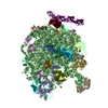
 Movie
Movie Controller

[English] 日本語
 Yorodumi
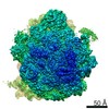
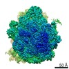
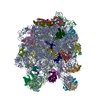
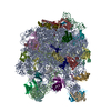
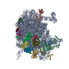
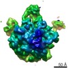
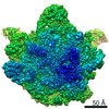
- PDB-6xz7: E. coli 50S ribosomal subunit in complex with dirithromycin, fMet... -
+ Open data

- Basic information
| Entry | Database: PDB / ID: 6xz7 | ||||||||||||||||||||||||||||||
|---|---|---|---|---|---|---|---|---|---|---|---|---|---|---|---|---|---|---|---|---|---|---|---|---|---|---|---|---|---|---|---|
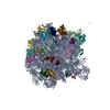
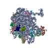
| Title | E. coli 50S ribosomal subunit in complex with dirithromycin, fMet-Phe-tRNA(Phe) and deacylated tRNA(iMet). | ||||||||||||||||||||||||||||||
 Components Components |
| ||||||||||||||||||||||||||||||
 Keywords Keywords | RIBOSOME / 50S ribosomal subunit / dirithromycin / antibiotics / cryo-EM | ||||||||||||||||||||||||||||||
| Function / homology |  Function and homology information Function and homology informationstringent response / translational termination / transcriptional attenuation / endoribonuclease inhibitor activity / positive regulation of ribosome biogenesis / RNA-binding transcription regulator activity / negative regulation of cytoplasmic translation / DnaA-L2 complex / translation repressor activity / negative regulation of DNA-templated DNA replication initiation ...stringent response / translational termination / transcriptional attenuation / endoribonuclease inhibitor activity / positive regulation of ribosome biogenesis / RNA-binding transcription regulator activity / negative regulation of cytoplasmic translation / DnaA-L2 complex / translation repressor activity / negative regulation of DNA-templated DNA replication initiation / mRNA regulatory element binding translation repressor activity / response to reactive oxygen species / cytosolic ribosome assembly / ribosome assembly / assembly of large subunit precursor of preribosome / regulation of cell growth / DNA-templated transcription termination / response to radiation / mRNA 5'-UTR binding / large ribosomal subunit / transferase activity / ribosome binding / 5S rRNA binding / ribosomal large subunit assembly / large ribosomal subunit rRNA binding / cytosolic large ribosomal subunit / cytoplasmic translation / tRNA binding / negative regulation of translation / rRNA binding / structural constituent of ribosome / ribosome / translation / response to antibiotic / negative regulation of DNA-templated transcription / mRNA binding / DNA binding / RNA binding / zinc ion binding / cytoplasm / cytosol Similarity search - Function | ||||||||||||||||||||||||||||||
| Biological species |   | ||||||||||||||||||||||||||||||
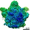
| Method | ELECTRON MICROSCOPY / single particle reconstruction / cryo EM / Resolution: 2.1 Å | ||||||||||||||||||||||||||||||
 Authors Authors | Pichkur, E.B. / Polikanov, Y.S. / Myasnikov, A.G. / Konevega, A.L. | ||||||||||||||||||||||||||||||
| Funding support |  Russian Federation, Russian Federation,  United States, 5items United States, 5items
| ||||||||||||||||||||||||||||||
 Citation Citation | Journal: RNA / Year: 2020 Title: Insights into the improved macrolide inhibitory activity from the high-resolution cryo-EM structure of dirithromycin bound to the 70S ribosome. Authors: Evgeny B Pichkur / Alena Paleskava / Andrey G Tereshchenkov / Pavel Kasatsky / Ekaterina S Komarova / Dmitrii I Shiriaev / Alexey A Bogdanov / Olga A Dontsova / Ilya A Osterman / Petr V ...Authors: Evgeny B Pichkur / Alena Paleskava / Andrey G Tereshchenkov / Pavel Kasatsky / Ekaterina S Komarova / Dmitrii I Shiriaev / Alexey A Bogdanov / Olga A Dontsova / Ilya A Osterman / Petr V Sergiev / Yury S Polikanov / Alexander G Myasnikov / Andrey L Konevega /  Abstract: Macrolides are one of the most successful and widely used classes of antibacterials, which kill or stop the growth of pathogenic bacteria by binding near the active site of the ribosome and ...Macrolides are one of the most successful and widely used classes of antibacterials, which kill or stop the growth of pathogenic bacteria by binding near the active site of the ribosome and interfering with protein synthesis. Dirithromycin is a derivative of the prototype macrolide erythromycin with additional hydrophobic side chain. In our recent study, we have discovered that the side chain of dirithromycin forms lone pair-π stacking interaction with the aromatic imidazole ring of the His69 residue in ribosomal protein uL4 of the 70S ribosome. In the current work, we found that neither the presence of the side chain, nor the additional contact with the ribosome, improve the binding affinity of dirithromycin to the ribosome. Nevertheless, we found that dirithromycin is a more potent inhibitor of in vitro protein synthesis in comparison with its parent compound, erythromycin. Using high-resolution cryo-electron microscopy, we determined the structure of the dirithromycin bound to the translating 70S ribosome, which suggests that the better inhibitory properties of the drug could be rationalized by the side chain of dirithromycin pointing into the lumen of the nascent peptide exit tunnel, where it can interfere with the normal passage of the growing polypeptide chain. | ||||||||||||||||||||||||||||||
| History |
|
- Structure visualization
| Movie |
Movie viewer |
|---|---|
| Structure viewer | Molecule: MolmilJmol/JSmol |
- Downloads & links
-Download
| PDBx/mmCIF format | 6xz7.cif.gz | 2.1 MB | Display | PDBx/mmCIF format |
|---|---|---|---|---|
| PDB format | pdb6xz7.ent.gz | 1.6 MB | Display | PDB format |
| PDBx/mmJSON format | 6xz7.json.gz | Tree view | PDBx/mmJSON format | |
| Others |  Other downloads Other downloads |
-Validation report
| Arichive directory | https://data.pdbj.org/pub/pdb/validation_reports/xz/6xz7ftp://data.pdbj.org/pub/pdb/validation_reports/xz/6xz7 | HTTPS FTP |
|---|
-Related structure data
| Related structure data | 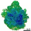 10655MC  6xzaC  6xzbC M: map data used to model this data C: citing same article ( |
|---|---|
| Similar structure data |

-Links
 PDBj
PDBj


- Assembly
| Deposited unit | 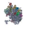
|
|---|---|
| 1 |
|
-Components
-RNA chain , 4 types, 4 molecules ABfg
| #1: RNA chain | Mass: 939609.250 Da / Num. of mol.: 1 / Source method: isolated from a natural source / Source: (natural) |
|---|---|
| #2: RNA chain | Mass: 38790.090 Da / Num. of mol.: 1 / Source method: isolated from a natural source / Source: (natural) |
| #32: RNA chain | Mass: 24541.719 Da / Num. of mol.: 1 / Source method: isolated from a natural source / Source: (natural) |
| #33: RNA chain | Mass: 25167.506 Da / Num. of mol.: 1 / Source method: isolated from a natural source / Source: (natural) |
+50S ribosomal protein ... , 29 types, 29 molecules CDEFGHIJKLMNOPQRSTUVWXYZabcde
-Non-polymers , 3 types, 707 molecules 


| #34: Chemical | ChemComp-MG /  Mass: 24.305 Da / Num. of mol.: 178 / Source method: obtained synthetically / Formula: Mg Mass: 24.305 Da / Num. of mol.: 178 / Source method: obtained synthetically / Formula: Mg#35: Chemical | ChemComp-DI0 / |  Mass: 835.074 Da / Num. of mol.: 1 / Source method: obtained synthetically / Formula: C42H78N2O14 / Feature type: SUBJECT OF INVESTIGATION / Comment: antibiotic*YM Mass: 835.074 Da / Num. of mol.: 1 / Source method: obtained synthetically / Formula: C42H78N2O14 / Feature type: SUBJECT OF INVESTIGATION / Comment: antibiotic*YM#36: Water | ChemComp-HOH / | Mass: 18.015 Da / Num. of mol.: 528 / Source method: isolated from a natural source / Formula: H2O |
|---|
-Details
| Has ligand of interest | Y |
|---|---|
| Has protein modification | Y |
-Experimental details
-Experiment
| Experiment | Method: ELECTRON MICROSCOPY |
|---|---|
| EM experiment | Aggregation state: PARTICLE / 3D reconstruction method: single particle reconstruction |
- Sample preparation
| Component |
| ||||||||||||||||||||||||
|---|---|---|---|---|---|---|---|---|---|---|---|---|---|---|---|---|---|---|---|---|---|---|---|---|---|
| Molecular weight | Value: 2.7 MDa / Experimental value: NO | ||||||||||||||||||||||||
| Source (natural) |
| ||||||||||||||||||||||||
| Buffer solution | pH: 7.5 | ||||||||||||||||||||||||
| Specimen | Embedding applied: NO / Shadowing applied: NO / Staining applied: NO / Vitrification applied: YES | ||||||||||||||||||||||||
| Specimen support | Grid material: COPPER/RHODIUM / Grid mesh size: 300 divisions/in. / Grid type: Quantifoil R2/2 | ||||||||||||||||||||||||
| Vitrification | Cryogen name: ETHANE |
- Electron microscopy imaging
| Experimental equipment |  Model: Titan Krios / Image courtesy: FEI Company |
|---|---|
| Microscopy | Model: FEI TITAN KRIOS |
| Electron gun | Electron source:  FIELD EMISSION GUN / Accelerating voltage: 300 kV / Illumination mode: SPOT SCAN FIELD EMISSION GUN / Accelerating voltage: 300 kV / Illumination mode: SPOT SCAN |
| Electron lens | Mode: BRIGHT FIELD / Nominal magnification: 75000 X / Nominal defocus max: 2200 nm / Nominal defocus min: 300 nm / Calibrated defocus min: 300 nm / Calibrated defocus max: 2200 nm / Cs: 0.1 mm / C2 aperture diameter: 100 µm / Alignment procedure: ZEMLIN TABLEAU |
| Specimen holder | Cryogen: NITROGEN |
| Image recording | Average exposure time: 1.4 sec. / Electron dose: 80 e/Å2 / Detector mode: INTEGRATING / Film or detector model: FEI FALCON II (4k x 4k) |
| Image scans | Movie frames/image: 28 / Used frames/image: 2-27 |
- Processing
| EM software |
| |||||||||||||||||||||||||||||||||||
|---|---|---|---|---|---|---|---|---|---|---|---|---|---|---|---|---|---|---|---|---|---|---|---|---|---|---|---|---|---|---|---|---|---|---|---|---|
| CTF correction | Type: PHASE FLIPPING AND AMPLITUDE CORRECTION | |||||||||||||||||||||||||||||||||||
| Particle selection | Num. of particles selected: 973000 | |||||||||||||||||||||||||||||||||||
| Symmetry | Point symmetry: C1 (asymmetric) | |||||||||||||||||||||||||||||||||||
| 3D reconstruction | Resolution: 2.1 Å / Resolution method: FSC 0.143 CUT-OFF / Num. of particles: 401905 / Symmetry type: POINT | |||||||||||||||||||||||||||||||||||
| Atomic model building | Protocol: OTHER / Space: REAL | |||||||||||||||||||||||||||||||||||
| Atomic model building | PDB-ID: 4YBB Accession code: 4YBB / Source name: PDB / Type: experimental model |

