Movie
Movie Controller

[English] 日本語
 Yorodumi
- PDB-6wts: CryoEM structure of the C. sordellii lethal toxin TcsL in complex... -
+ Open data

- Basic information
| Entry | Database: PDB / ID: 6wts | ||||||||||||
|---|---|---|---|---|---|---|---|---|---|---|---|---|---|
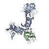
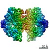
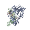
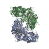
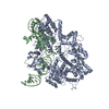
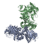
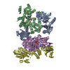
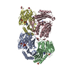
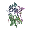
| Title | CryoEM structure of the C. sordellii lethal toxin TcsL in complex with SEMA6A | ||||||||||||
 Components Components |
| ||||||||||||
 Keywords Keywords | SIGNALING PROTEIN/TOXIN / cryoEM / signaling protein-toxin complex | ||||||||||||
| Function / homology |  Function and homology information Function and homology informationnegative regulation of cell adhesion involved in sprouting angiogenesis / negative regulation of sprouting angiogenesis / semaphorin receptor binding / Other semaphorin interactions / negative regulation of vascular endothelial growth factor signaling pathway / negative regulation of axon extension involved in axon guidance / positive regulation of neuron migration / chemorepellent activity / neural crest cell migration / host cell cytosol ...negative regulation of cell adhesion involved in sprouting angiogenesis / negative regulation of sprouting angiogenesis / semaphorin receptor binding / Other semaphorin interactions / negative regulation of vascular endothelial growth factor signaling pathway / negative regulation of axon extension involved in axon guidance / positive regulation of neuron migration / chemorepellent activity / neural crest cell migration / host cell cytosol / Transferases; Glycosyltransferases; Hexosyltransferases / negative chemotaxis / glycosyltransferase activity / semaphorin-plexin signaling pathway / cellular response to vascular endothelial growth factor stimulus / cysteine-type peptidase activity / cytoskeleton organization / negative regulation of angiogenesis / host cell endosome membrane / axon guidance / animal organ morphogenesis / negative regulation of ERK1 and ERK2 cascade / nervous system development / toxin activity / cell surface receptor signaling pathway / positive regulation of cell migration / axon / apoptotic process / host cell plasma membrane / extracellular space / extracellular region / membrane / metal ion binding / plasma membrane Similarity search - Function | ||||||||||||
| Biological species |  Homo sapiens (human) Homo sapiens (human) Paeniclostridium sordellii (bacteria) Paeniclostridium sordellii (bacteria) | ||||||||||||
| Method | ELECTRON MICROSCOPY / single particle reconstruction / cryo EM / Resolution: 3.3 Å | ||||||||||||
 Authors Authors | Kucharska, I. / Rubinstein, J.L. / Julien, J.P. | ||||||||||||
| Funding support |  Canada, 3items Canada, 3items
| ||||||||||||
 Citation Citation | Journal: Cell / Year: 2020 Title: Recognition of Semaphorin Proteins by P. sordellii Lethal Toxin Reveals Principles of Receptor Specificity in Clostridial Toxins. Authors: Hunsang Lee / Greg L Beilhartz / Iga Kucharska / Swetha Raman / Hong Cui / Mandy Hiu Yi Lam / Huazhu Liang / John L Rubinstein / Daniel Schramek / Jean-Philippe Julien / Roman A Melnyk / Mikko Taipale / Abstract: Pathogenic clostridial species secrete potent toxins that induce severe host tissue damage. Paeniclostridium sordellii lethal toxin (TcsL) causes an almost invariably lethal toxic shock syndrome ...Pathogenic clostridial species secrete potent toxins that induce severe host tissue damage. Paeniclostridium sordellii lethal toxin (TcsL) causes an almost invariably lethal toxic shock syndrome associated with gynecological infections. TcsL is 87% similar to C. difficile TcdB, which enters host cells via Frizzled receptors in colon epithelium. However, P. sordellii infections target vascular endothelium, suggesting that TcsL exploits another receptor. Here, using CRISPR/Cas9 screening, we establish semaphorins SEMA6A and SEMA6B as TcsL receptors. We demonstrate that recombinant SEMA6A can protect mice from TcsL-induced edema. A 3.3 Å cryo-EM structure shows that TcsL binds SEMA6A with the same region that in TcdB binds structurally unrelated Frizzled. Remarkably, 15 mutations in this evolutionarily divergent surface are sufficient to switch binding specificity of TcsL to that of TcdB. Our findings establish semaphorins as physiologically relevant receptors for TcsL and reveal the molecular basis for the difference in tissue targeting and disease pathogenesis between highly related toxins. | ||||||||||||
| History |
|
- Structure visualization
| Movie |
Movie viewer |
|---|---|
| Structure viewer | Molecule: MolmilJmol/JSmol |
- Downloads & links
-Download
| PDBx/mmCIF format | 6wts.cif.gz | 241.3 KB | Display | PDBx/mmCIF format |
|---|---|---|---|---|
| PDB format | pdb6wts.ent.gz | 175.5 KB | Display | PDB format |
| PDBx/mmJSON format | 6wts.json.gz | Tree view | PDBx/mmJSON format | |
| Others |  Other downloads Other downloads |
-Validation report
| Summary document | 6wts_validation.pdf.gz | 1 MB | Display | wwPDB validaton report |
|---|---|---|---|---|
| Full document | 6wts_full_validation.pdf.gz | 1 MB | Display | |
| Data in XML | 6wts_validation.xml.gz | 37.2 KB | Display | |
| Data in CIF | 6wts_validation.cif.gz | 56.3 KB | Display | |
| Arichive directory | https://data.pdbj.org/pub/pdb/validation_reports/wt/6wtsftp://data.pdbj.org/pub/pdb/validation_reports/wt/6wts | HTTPS FTP |
-Related structure data
| Related structure data | 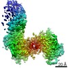 21898MC M: map data used to model this data C: citing same article ( |
|---|---|
| Similar structure data |
-Links
 PDBj
PDBj


- Assembly
| Deposited unit | 
|
|---|---|
| 1 |
|
-Components
| #1: Protein | Mass: 63857.410 Da / Num. of mol.: 2 Source method: isolated from a genetically manipulated source Source: (gene. exp.) Homo sapiens (human) / Gene: SEMA6A, KIAA1368, SEMAQ / Production host: Homo sapiens (human) / References: UniProt: Q9H2E6#2: Protein | | Mass: 63315.875 Da / Num. of mol.: 1 / Fragment: UNP residues 1285-1804 Source method: isolated from a genetically manipulated source Source: (gene. exp.) Paeniclostridium sordellii (bacteria) / Gene: tcsL, JGS6382_PCS1300041 / Production host: References: UniProt: V5T923, Transferases; Glycosyltransferases; Hexosyltransferases #3: Polysaccharide | Source method: isolated from a genetically manipulated source #4: Sugar | ChemComp-NAG /   Type: D-saccharide, beta linking / Mass: 221.208 Da / Num. of mol.: 4 Type: D-saccharide, beta linking / Mass: 221.208 Da / Num. of mol.: 4Source method: isolated from a genetically manipulated source Formula: C8H15NO6 Has ligand of interest | N | Has protein modification | Y | |
|---|
-Experimental details
-Experiment
| Experiment | Method: ELECTRON MICROSCOPY |
|---|---|
| EM experiment | Aggregation state: PARTICLE / 3D reconstruction method: single particle reconstruction |
- Sample preparation
| Component |
| ||||||||||||||||||||||||
|---|---|---|---|---|---|---|---|---|---|---|---|---|---|---|---|---|---|---|---|---|---|---|---|---|---|
| Source (natural) |
| ||||||||||||||||||||||||
| Source (recombinant) |
| ||||||||||||||||||||||||
| Buffer solution | pH: 7 | ||||||||||||||||||||||||
| Specimen | Conc.: 0.9 mg/ml / Embedding applied: NO / Shadowing applied: NO / Staining applied: NO / Vitrification applied: YES | ||||||||||||||||||||||||
| Vitrification | Cryogen name: ETHANE-PROPANE |
- Electron microscopy imaging
| Experimental equipment |  Model: Titan Krios / Image courtesy: FEI Company |
|---|---|
| Microscopy | Model: FEI TITAN KRIOS |
| Electron gun | Electron source:  FIELD EMISSION GUN / Accelerating voltage: 300 kV / Illumination mode: FLOOD BEAM FIELD EMISSION GUN / Accelerating voltage: 300 kV / Illumination mode: FLOOD BEAM |
| Electron lens | Mode: BRIGHT FIELD |
| Image recording | Electron dose: 45.24 e/Å2 / Film or detector model: OTHER / Details: Dataset was collected with FEI Falcon IV. |
- Processing
| Software | Name: PHENIX / Version: 1.18rc5_3822: / Classification: refinement | ||||||||||||||||||||||||
|---|---|---|---|---|---|---|---|---|---|---|---|---|---|---|---|---|---|---|---|---|---|---|---|---|---|
| EM software |
| ||||||||||||||||||||||||
| CTF correction | Type: PHASE FLIPPING AND AMPLITUDE CORRECTION | ||||||||||||||||||||||||
| Symmetry | Point symmetry: C1 (asymmetric) | ||||||||||||||||||||||||
| 3D reconstruction | Resolution: 3.3 Å / Resolution method: FSC 0.143 CUT-OFF / Num. of particles: 179188 / Symmetry type: POINT | ||||||||||||||||||||||||
| Atomic model building | PDB-ID: 6C0B Accession code: 6C0B / Source name: PDB / Type: experimental model | ||||||||||||||||||||||||
| Refinement | Cross valid method: NONE Stereochemistry target values: GeoStd + Monomer Library + CDL v1.2 | ||||||||||||||||||||||||
| Displacement parameters | Biso mean: 76.11 Å2 | ||||||||||||||||||||||||
| Refine LS restraints |
|