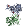
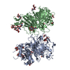
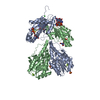
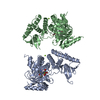
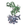
Journal: Cell / Year: 2020 Title: Recognition of Semaphorin Proteins by P. sordellii Lethal Toxin Reveals Principles of Receptor Specificity in Clostridial Toxins. Authors: Hunsang Lee / Greg L Beilhartz / Iga Kucharska / Swetha Raman / Hong Cui / Mandy Hiu Yi Lam / Huazhu Liang / John L Rubinstein / Daniel Schramek / Jean-Philippe Julien / Roman A Melnyk / Mikko Taipale / Abstract: Pathogenic clostridial species secrete potent toxins that induce severe host tissue damage. Paeniclostridium sordellii lethal toxin (TcsL) causes an almost invariably lethal toxic shock syndrome ...Pathogenic clostridial species secrete potent toxins that induce severe host tissue damage. Paeniclostridium sordellii lethal toxin (TcsL) causes an almost invariably lethal toxic shock syndrome associated with gynecological infections. TcsL is 87% similar to C. difficile TcdB, which enters host cells via Frizzled receptors in colon epithelium. However, P. sordellii infections target vascular endothelium, suggesting that TcsL exploits another receptor. Here, using CRISPR/Cas9 screening, we establish semaphorins SEMA6A and SEMA6B as TcsL receptors. We demonstrate that recombinant SEMA6A can protect mice from TcsL-induced edema. A 3.3 Å cryo-EM structure shows that TcsL binds SEMA6A with the same region that in TcdB binds structurally unrelated Frizzled. Remarkably, 15 mutations in this evolutionarily divergent surface are sufficient to switch binding specificity of TcsL to that of TcdB. Our findings establish semaphorins as physiologically relevant receptors for TcsL and reveal the molecular basis for the difference in tissue targeting and disease pathogenesis between highly related toxins.
History
Deposition
May 3, 2020
-
Header (metadata) release
Jul 8, 2020
-
Map release
Jul 8, 2020
-
Update
Jul 8, 2020
-
Current status
Jul 8, 2020
Processing site: RCSB / Status: Released
-
Structure visualization
Movie
Surface view with section colored by density value
In the structure databanks used in Yorodumi, some data are registered as the other names, "COVID-19 virus" and "2019-nCoV". Here are the details of the virus and the list of structure data.
Jan 31, 2019. EMDB accession codes are about to change! (news from PDBe EMDB page)
EMDB accession codes are about to change! (news from PDBe EMDB page)
The allocation of 4 digits for EMDB accession codes will soon come to an end. Whilst these codes will remain in use, new EMDB accession codes will include an additional digit and will expand incrementally as the available range of codes is exhausted. The current 4-digit format prefixed with “EMD-” (i.e. EMD-XXXX) will advance to a 5-digit format (i.e. EMD-XXXXX), and so on. It is currently estimated that the 4-digit codes will be depleted around Spring 2019, at which point the 5-digit format will come into force.
The EM Navigator/Yorodumi systems omit the EMD- prefix.
Related info.:Q: What is EMD? / ID/Accession-code notation in Yorodumi/EM Navigator
Yorodumi is a browser for structure data from EMDB, PDB, SASBDB, etc.
This page is also the successor to EM Navigator detail page, and also detail information page/front-end page for Omokage search.
The word "yorodu" (or yorozu) is an old Japanese word meaning "ten thousand". "mi" (miru) is to see.
Related info.:EMDB / PDB / SASBDB / Comparison of 3 databanks / Yorodumi Search / Aug 31, 2016. New EM Navigator & Yorodumi / Yorodumi Papers / Jmol/JSmol / Function and homology information / Changes in new EM Navigator and Yorodumi
 Movie
Movie Controller


 Open data
 Basic information
 Map data
 Sample
 Function and homology information
 Homo sapiens (human)
 Authors
 Canada, 3 items
  Citation
 Structure visualization
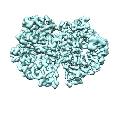
 Downloads & links
 emd_21899.png
 http://ftp.pdbj.org/pub/emdb/structures/EMD-21899
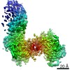


 X (Sec.)
 Y (Row.)
 Z (Col.)


 Sample components
 Processing
 Electron microscopy
 FIELD EMISSION GUN

