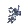
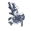
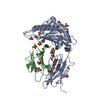
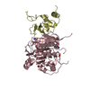
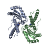
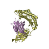
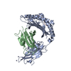
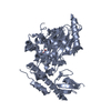
[English] 日本語
Yorodumi
- PDB-2xue: CRYSTAL STRUCTURE OF JMJD3 -

+
Open data


ID or keywords:

Loading...

-
Basic information

Entry
Database: PDB / ID: 2xue
TitleCRYSTAL STRUCTURE OF JMJD3
ComponentsLYSINE-SPECIFIC DEMETHYLASE 6B
KeywordsOXIDOREDUCTASE / HISTONE METHYLASE
Function / homology
Function and homology information


[histone H3]-trimethyl-L-lysine27 demethylase / histone H3K27me2/H3K27me3 demethylase activity / endothelial cell differentiation / cardiac muscle cell differentiation / MLL3/4 complex / mesodermal cell differentiation / inflammatory response to antigenic stimulus / histone demethylase activity / cell fate commitment / response to fungicide ...[histone H3]-trimethyl-L-lysine27 demethylase / histone H3K27me2/H3K27me3 demethylase activity / endothelial cell differentiation / cardiac muscle cell differentiation / MLL3/4 complex / mesodermal cell differentiation / inflammatory response to antigenic stimulus / histone demethylase activity / cell fate commitment / response to fungicide / Chromatin modifications during the maternal to zygotic transition (MZT) / response to activity / hippocampus development / HDMs demethylate histones / chromatin DNA binding / beta-catenin binding / cellular response to hydrogen peroxide / positive regulation of cold-induced thermogenesis / regulation of gene expression / Oxidative Stress Induced Senescence / chromatin remodeling / RNA polymerase II cis-regulatory region sequence-specific DNA binding / positive regulation of transcription by RNA polymerase II / nucleoplasm / nucleus / metal ion binding
Similarity search - Function
Methane Monooxygenase Hydroxylase; Chain G, domain 1 - #1370 / Cysteine Rich Protein - #20 / : / : / : / Lysine-specific demethylase 6/UTY, C-terminal helical domain / KDM6, GATA-like / Cysteine Rich Protein / Cupin / JmjC domain, hydroxylase ...Methane Monooxygenase Hydroxylase; Chain G, domain 1 - #1370 / Cysteine Rich Protein - #20 / : / : / : / Lysine-specific demethylase 6/UTY, C-terminal helical domain / KDM6, GATA-like / Cysteine Rich Protein / Cupin / JmjC domain, hydroxylase / A domain family that is part of the cupin metalloenzyme superfamily. / JmjC domain / JmjC domain profile. / Methane Monooxygenase Hydroxylase; Chain G, domain 1 / Ribbon / Jelly Rolls / Up-down Bundle / Sandwich / Mainly Beta / Mainly Alpha
Similarity search - Domain/homology
2-OXOGLUTARIC ACID / : / Lysine-specific demethylase 6B
Similarity search - Component
Biological speciesHOMO SAPIENS (human)
MethodX-RAY DIFFRACTION / SYNCHROTRON / SAD / Resolution: 2 Å
AuthorsChung, C. / Rowland, P. / Mosley, J. / Thomas, P.J.
CitationJournal: Nature / Year: 2012
Title: A Selective Jumonji H3K27 Demethylase Inhibitor Modulates the Proinflammatory Macrophage Response
Authors: Kruidenier, L. / Chung, C. / Cheng, Z. / Liddle, J. / Che, K. / Joberty, G. / Bantscheff, M. / Bountra, C. / Bridges, A. / Diallo, H. / Eberhard, D. / Hutchinson, S. / Jones, E. / Katso, R. ...Authors: Kruidenier, L. / Chung, C. / Cheng, Z. / Liddle, J. / Che, K. / Joberty, G. / Bantscheff, M. / Bountra, C. / Bridges, A. / Diallo, H. / Eberhard, D. / Hutchinson, S. / Jones, E. / Katso, R. / Leveridge, M. / Mander, P.K. / Mosley, J. / Ramirez-Molina, C. / Rowland, P. / Schofield, C.J. / Sheppard, R.J. / Smith, J.E. / Swales, C. / Tanner, R. / Thomas, P. / Tumber, A. / Drewes, G. / Oppermann, U. / Patel, D.J. / Lee, K. / Wilson, D.M.
History
DepositionOct 19, 2010Deposition site: PDBE / Processing site: PDBE
Revision 1.0Dec 28, 2011Provider: repository / Type: Initial release
Revision 1.1Jul 25, 2012Group: Database references
Revision 1.2Aug 29, 2012Group: Database references
Revision 1.3May 8, 2024Group: Data collection / Database references ...Data collection / Database references / Derived calculations / Other
Category: chem_comp_atom / chem_comp_bond ...chem_comp_atom / chem_comp_bond / database_2 / pdbx_database_status / pdbx_struct_conn_angle / struct_conn / struct_site
Item: _database_2.pdbx_DOI / _database_2.pdbx_database_accession ..._database_2.pdbx_DOI / _database_2.pdbx_database_accession / _pdbx_database_status.status_code_sf / _pdbx_struct_conn_angle.ptnr1_auth_comp_id / _pdbx_struct_conn_angle.ptnr1_auth_seq_id / _pdbx_struct_conn_angle.ptnr1_label_asym_id / _pdbx_struct_conn_angle.ptnr1_label_atom_id / _pdbx_struct_conn_angle.ptnr1_label_comp_id / _pdbx_struct_conn_angle.ptnr1_label_seq_id / _pdbx_struct_conn_angle.ptnr2_auth_comp_id / _pdbx_struct_conn_angle.ptnr2_auth_seq_id / _pdbx_struct_conn_angle.ptnr2_label_asym_id / _pdbx_struct_conn_angle.ptnr2_label_atom_id / _pdbx_struct_conn_angle.ptnr2_label_comp_id / _pdbx_struct_conn_angle.ptnr3_auth_comp_id / _pdbx_struct_conn_angle.ptnr3_auth_seq_id / _pdbx_struct_conn_angle.ptnr3_label_asym_id / _pdbx_struct_conn_angle.ptnr3_label_atom_id / _pdbx_struct_conn_angle.ptnr3_label_comp_id / _pdbx_struct_conn_angle.ptnr3_label_seq_id / _pdbx_struct_conn_angle.value / _struct_conn.pdbx_dist_value / _struct_conn.ptnr1_auth_comp_id / _struct_conn.ptnr1_auth_seq_id / _struct_conn.ptnr1_label_asym_id / _struct_conn.ptnr1_label_atom_id / _struct_conn.ptnr1_label_comp_id / _struct_conn.ptnr1_label_seq_id / _struct_conn.ptnr2_auth_comp_id / _struct_conn.ptnr2_auth_seq_id / _struct_conn.ptnr2_label_asym_id / _struct_conn.ptnr2_label_atom_id / _struct_conn.ptnr2_label_comp_id / _struct_conn.ptnr2_label_seq_id / _struct_site.pdbx_auth_asym_id / _struct_site.pdbx_auth_comp_id / _struct_site.pdbx_auth_seq_id

-
Structure visualization

Structure viewerMolecule:
MolmilJmol/JSmol

Downloads & links

-
Assembly

Deposited unit
A: LYSINE-SPECIFIC DEMETHYLASE 6B
B: LYSINE-SPECIFIC DEMETHYLASE 6B
hetero molecules


Theoretical massNumber of molelcules
Total (without water)116,56510
Polymers115,8382
Non-polymers7278
Water10,088560
1
A: LYSINE-SPECIFIC DEMETHYLASE 6B
hetero molecules


Theoretical massNumber of molelcules
Total (without water)58,2835
Polymers57,9191
Non-polymers3634
Water181
TypeNameSymmetry operationNumber
identity operation1_555x,y,z1
MethodPISA
2
B: LYSINE-SPECIFIC DEMETHYLASE 6B
hetero molecules


Theoretical massNumber of molelcules
Total (without water)58,2835
Polymers57,9191
Non-polymers3634
Water181
TypeNameSymmetry operationNumber
identity operation1_555x,y,z1
MethodPISA
Unit cell
Length a, b, c (Å)61.215, 65.154, 77.462
Angle α, β, γ (deg.)86.09, 67.19, 68.26
Int Tables number1
Space group name H-MP1

-
Components

-
Protein , 1 types, 2 molecules AB

#1: Protein LYSINE-SPECIFIC DEMETHYLASE 6B / JMJD3 / JMJC DOMAIN-CONTAINING PROTEIN 3 / JUMONJI DOMAIN-CONTAINING PROTEIN 3 / LYSINE DEMETHYLASE 6B


Mass: 57919.098 Da / Num. of mol.: 2 / Fragment: CATALYTIC DOMAIN, RESIDUES 1138-1640
Source method: isolated from a genetically manipulated source
Source: (gene. exp.) HOMO SAPIENS (human) / Cell line (production host): SF9 / Production host: SPODOPTERA FRUGIPERDA (fall armyworm)
References: UniProt: O15054, Oxidoreductases; Acting on paired donors, with incorporation or reduction of molecular oxygen; With 2-oxoglutarate as one donor, and incorporation of one atom of oxygen into each donor

-
Non-polymers , 5 types, 568 molecules

#2: Chemical ChemComp-ZN / ZINC ION


Mass: 65.409 Da / Num. of mol.: 2 / Source method: obtained synthetically / Formula: Zn
#3: Chemical ChemComp-SO4 / SULFATE ION


Mass: 96.063 Da / Num. of mol.: 2 / Source method: obtained synthetically / Formula: SO4
#4: Chemical ChemComp-FE / FE (III) ION


Mass: 55.845 Da / Num. of mol.: 2 / Source method: obtained synthetically / Formula: Fe
#5: Chemical ChemComp-AKG / 2-OXOGLUTARIC ACID


Mass: 146.098 Da / Num. of mol.: 2 / Source method: obtained synthetically / Formula: C5H6O5
#6: Water ChemComp-HOH / water


Mass: 18.015 Da / Num. of mol.: 560 / Source method: isolated from a natural source / Formula: H2O

-
Experimental details

-
Experiment

ExperimentMethod: X-RAY DIFFRACTION / Number of used crystals: 1

-
Sample preparation

CrystalDensity Matthews: 2.53 Å3/Da / Density % sol: 51.04 % / Description: NONE
Crystal growpH: 8.5
Details: MORPHEUS SCREEN (MOLECULAR DIMENSIONS) CONDITION A12 (0.03M MGCL2, 0.03M CACL2, 0.1M TRIS HCL/BICINE PH 8.5, 12.5%V/V MPD, 12.5%W/V PEG1000, 12.5%W/V PEG3350)

-
Data collection

Diffraction
IDMean temperature (K)Crystal-ID
11001
21
Diffraction source
SourceSiteBeamlineIDWavelength
SYNCHROTRONESRF ID14-410.981
SYNCHROTRONESRF ID14-420.9795
Detector
TypeIDDetectorDate
ADSC CCD1CCDApr 28, 2010
ADSC CCD2CCD
Radiation
IDProtocolMonochromatic (M) / Laue (L)Scattering typeWavelength-ID
1SINGLE WAVELENGTHMx-ray1
2Mx-ray1
Radiation wavelength
IDWavelength (Å)Relative weight
10.9811
20.97951
ReflectionResolution: 2→40 Å / Num. obs: 61840 / % possible obs: 89.9 % / Observed criterion σ(I): 2 / Redundancy: 2.3 % / Rmerge(I) obs: 0.06 / Net I/σ(I): 12.3
Reflection shellResolution: 2→2.03 Å / Redundancy: 2.1 % / Rmerge(I) obs: 0.51 / Mean I/σ(I) obs: 1.5 / % possible all: 56.1

-
Processing

Software
NameVersionClassification
DENZOdata reduction
SCALEPACKdata scaling
autoSHARPphasing
RESOLVEphasing
REFMAC5.5.0109refinement
RefinementMethod to determine structure: SAD
Starting model: NONE

Resolution: 2→71.13 Å / Cor.coef. Fo:Fc: 0.962 / Cor.coef. Fo:Fc free: 0.945 / SU B: 10.488 / SU ML: 0.125 / Cross valid method: THROUGHOUT / ESU R: 0.189 / ESU R Free: 0.161 / Stereochemistry target values: MAXIMUM LIKELIHOOD
Details: HYDROGENS HAVE BEEN ADDED IN THE RIDING POSITIONS. ATOM RECORD CONTAINS SUM OF TLS AND RESIDUAL B FACTORS. ANISOU RECORD CONTAINS SUM OF TLS AND RESIDUAL U FACTORS.
RfactorNum. reflection% reflectionSelection details
Rfree0.21574 2450 4 %RANDOM
Rwork0.17932 ---
obs0.18081 59387 89.76 %-
Solvent computationIon probe radii: 0.8 Å / Shrinkage radii: 0.8 Å / VDW probe radii: 1.4 Å / Solvent model: BABINET MODEL WITH MASK
Displacement parametersBiso mean: 44.196 Å2
Baniso -1Baniso -2Baniso -3
1-0.95 Å2-2.46 Å2-1.11 Å2
2--1.4 Å21.85 Å2
3---0.08 Å2
Refinement stepCycle: LAST / Resolution: 2→71.13 Å
ProteinNucleic acidLigandSolventTotal
Num. atoms6805 0 34 560 7399
Refine LS restraints
Refine-IDTypeDev idealDev ideal targetNumber
X-RAY DIFFRACTIONr_bond_refined_d0.010.0227068
X-RAY DIFFRACTIONr_bond_other_d0.0010.024722
X-RAY DIFFRACTIONr_angle_refined_deg1.0891.9359612
X-RAY DIFFRACTIONr_angle_other_deg0.8063.00111455
X-RAY DIFFRACTIONr_dihedral_angle_1_deg7.4855852
X-RAY DIFFRACTIONr_dihedral_angle_2_deg31.90123.761335
X-RAY DIFFRACTIONr_dihedral_angle_3_deg12.713151141
X-RAY DIFFRACTIONr_dihedral_angle_4_deg13.0191546
X-RAY DIFFRACTIONr_chiral_restr0.0630.21048
X-RAY DIFFRACTIONr_gen_planes_refined0.0060.0217852
X-RAY DIFFRACTIONr_gen_planes_other0.0010.021468
X-RAY DIFFRACTIONr_nbd_refined
X-RAY DIFFRACTIONr_nbd_other
X-RAY DIFFRACTIONr_nbtor_refined
X-RAY DIFFRACTIONr_nbtor_other
X-RAY DIFFRACTIONr_xyhbond_nbd_refined
X-RAY DIFFRACTIONr_xyhbond_nbd_other
X-RAY DIFFRACTIONr_metal_ion_refined
X-RAY DIFFRACTIONr_metal_ion_other
X-RAY DIFFRACTIONr_symmetry_vdw_refined
X-RAY DIFFRACTIONr_symmetry_vdw_other
X-RAY DIFFRACTIONr_symmetry_hbond_refined
X-RAY DIFFRACTIONr_symmetry_hbond_other
X-RAY DIFFRACTIONr_symmetry_metal_ion_refined
X-RAY DIFFRACTIONr_symmetry_metal_ion_other
X-RAY DIFFRACTIONr_mcbond_it0.87624262
X-RAY DIFFRACTIONr_mcbond_other0.09821712
X-RAY DIFFRACTIONr_mcangle_it1.72246917
X-RAY DIFFRACTIONr_mcangle_other
X-RAY DIFFRACTIONr_scbond_it2.02542806
X-RAY DIFFRACTIONr_scbond_other
X-RAY DIFFRACTIONr_scangle_it3.07262695
X-RAY DIFFRACTIONr_scangle_other
X-RAY DIFFRACTIONr_long_range_B_refined
X-RAY DIFFRACTIONr_long_range_B_other
X-RAY DIFFRACTIONr_rigid_bond_restr
X-RAY DIFFRACTIONr_sphericity_free
X-RAY DIFFRACTIONr_sphericity_bonded
LS refinement shellResolution: 2.001→2.053 Å / Total num. of bins used: 20
RfactorNum. reflection% reflection
Rfree0.348 103 -
Rwork0.29 2822 -
obs--57.31 %
Refinement TLS params.

Method: refined / Refine-ID: X-RAY DIFFRACTION

IDL112)L122)L132)L222)L232)L332)S11 (Å °)S12 (Å °)S13 (Å °)S21 (Å °)S22 (Å °)S23 (Å °)S31 (Å °)S32 (Å °)S33 (Å °)T112)T122)T132)T222)T232)T332)Origin x (Å)Origin y (Å)Origin z (Å)
10.6615-0.33220.42440.3081-0.32040.6222-0.0007-0.018-0.0178-0.00380.01150.0061-0.0018-0.0344-0.01070.0336-0.01470.00090.0185-0.01510.024957.160117.094-13.7037
20.594-0.24170.22470.4552-0.27520.5281-0.01080.0028-0.0064-0.03680.0224-0.03260.0502-0.0428-0.01160.0355-0.01030.01160.0116-0.01110.023574.702745.692319.3965
Refinement TLS group
IDRefine-IDRefine TLS-IDAuth asym-IDAuth seq-ID
1X-RAY DIFFRACTION1A1178 - 4000
2X-RAY DIFFRACTION2B1181 - 4000

+
About Yorodumi

-
News

-
Feb 9, 2022. New format data for meta-information of EMDB entries

New format data for meta-information of EMDB entries

  • Version 3 of the EMDB header file is now the official format.
  • The previous official version 1.9 will be removed from the archive.

Related info.:EMDB header

External links:wwPDB to switch to version 3 of the EMDB data model

-
Aug 12, 2020. Covid-19 info

Covid-19 info

URL: https://pdbjlvh1.pdbj.org/emnavi/covid19.php

New page: Covid-19 featured information page in EM Navigator.

Related info.:Covid-19 info / Mar 5, 2020. Novel coronavirus structure data

+
Mar 5, 2020. Novel coronavirus structure data

Novel coronavirus structure data

Related info.:Yorodumi Speices / Aug 12, 2020. Covid-19 info

External links:COVID-19 featured content - PDBj / Molecule of the Month (242):Coronavirus Proteases

+
Jan 31, 2019. EMDB accession codes are about to change! (news from PDBe EMDB page)

EMDB accession codes are about to change! (news from PDBe EMDB page)

  • The allocation of 4 digits for EMDB accession codes will soon come to an end. Whilst these codes will remain in use, new EMDB accession codes will include an additional digit and will expand incrementally as the available range of codes is exhausted. The current 4-digit format prefixed with “EMD-” (i.e. EMD-XXXX) will advance to a 5-digit format (i.e. EMD-XXXXX), and so on. It is currently estimated that the 4-digit codes will be depleted around Spring 2019, at which point the 5-digit format will come into force.
  • The EM Navigator/Yorodumi systems omit the EMD- prefix.

Related info.:Q: What is EMD? / ID/Accession-code notation in Yorodumi/EM Navigator

External links:EMDB Accession Codes are Changing Soon! / Contact to PDBj

+
Jul 12, 2017. Major update of PDB

Major update of PDB

  • wwPDB released updated PDB data conforming to the new PDBx/mmCIF dictionary.
  • This is a major update changing the version number from 4 to 5, and with Remediation, in which all the entries are updated.
  • In this update, many items about electron microscopy experimental information are reorganized (e.g. em_software).
  • Now, EM Navigator and Yorodumi are based on the updated data.

External links:wwPDB Remediation / Enriched Model Files Conforming to OneDep Data Standards Now Available in the PDB FTP Archive

-
Yorodumi

Thousand views of thousand structures

  • Yorodumi is a browser for structure data from EMDB, PDB, SASBDB, etc.
  • This page is also the successor to EM Navigator detail page, and also detail information page/front-end page for Omokage search.
  • The word "yorodu" (or yorozu) is an old Japanese word meaning "ten thousand". "mi" (miru) is to see.

Related info.:EMDB / PDB / SASBDB / Comparison of 3 databanks / Yorodumi Search / Aug 31, 2016. New EM Navigator & Yorodumi / Yorodumi Papers / Jmol/JSmol / Function and homology information / Changes in new EM Navigator and Yorodumi

Read more