 Movie
Movie Controller


+ Open data

- Basic information
| Entry | Database: PDB / ID: 6wb8 | |||||||||||||||||||||||||||||||||||||||||||||||||||||||||||||||
|---|---|---|---|---|---|---|---|---|---|---|---|---|---|---|---|---|---|---|---|---|---|---|---|---|---|---|---|---|---|---|---|---|---|---|---|---|---|---|---|---|---|---|---|---|---|---|---|---|---|---|---|---|---|---|---|---|---|---|---|---|---|---|---|---|
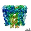
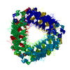
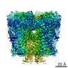
| Title | Cryo-EM structure of PKD2 C331S disease variant | |||||||||||||||||||||||||||||||||||||||||||||||||||||||||||||||
 Components Components | Polycystin-2 | |||||||||||||||||||||||||||||||||||||||||||||||||||||||||||||||
 Keywords Keywords | TRANSPORT PROTEIN / PKD2 | |||||||||||||||||||||||||||||||||||||||||||||||||||||||||||||||
| Function / homology |  Function and homology information Function and homology informationdetection of nodal flow / metanephric smooth muscle tissue development / metanephric cortex development / metanephric cortical collecting duct development / metanephric distal tubule development / polycystin complex / mesonephric tubule development / mesonephric duct development / metanephric part of ureteric bud development / renal tubule morphogenesis ...detection of nodal flow / metanephric smooth muscle tissue development / metanephric cortex development / metanephric cortical collecting duct development / metanephric distal tubule development / polycystin complex / mesonephric tubule development / mesonephric duct development / metanephric part of ureteric bud development / renal tubule morphogenesis / determination of liver left/right asymmetry / metanephric ascending thin limb development / metanephric mesenchyme development / metanephric S-shaped body morphogenesis / basal cortex / placenta blood vessel development / renal artery morphogenesis / HLH domain binding / VxPx cargo-targeting to cilium / cilium organization / migrasome / neural tube development / cellular response to fluid shear stress / muscle alpha-actinin binding / regulation of calcium ion import / calcium-induced calcium release activity / detection of mechanical stimulus / determination of left/right symmetry / voltage-gated monoatomic ion channel activity / embryonic placenta development / cellular response to hydrostatic pressure / aorta development / cation channel complex / branching involved in ureteric bud morphogenesis / non-motile cilium / outward rectifier potassium channel activity / motile cilium / actinin binding / cellular response to osmotic stress / negative regulation of G1/S transition of mitotic cell cycle / voltage-gated sodium channel activity / heart looping / spinal cord development / ciliary membrane / voltage-gated monoatomic cation channel activity / positive regulation of phospholipase C-activating G protein-coupled receptor signaling pathway / protein heterotetramerization / cytoplasmic side of endoplasmic reticulum membrane / potassium channel activity / voltage-gated potassium channel activity / centrosome duplication / transcription regulator inhibitor activity / cell surface receptor signaling pathway via JAK-STAT / voltage-gated calcium channel activity / release of sequestered calcium ion into cytosol / monoatomic cation channel activity / cytoskeletal protein binding / cellular response to calcium ion / cellular response to cAMP / liver development / potassium ion transmembrane transport / cytoplasmic vesicle membrane / basal plasma membrane / sodium ion transmembrane transport / lumenal side of endoplasmic reticulum membrane / cellular response to reactive oxygen species / protein tetramerization / phosphoprotein binding / Wnt signaling pathway / mitotic spindle / positive regulation of nitric oxide biosynthetic process / calcium ion transmembrane transport / heart development / calcium ion transport / transmembrane transport / cell-cell junction / cilium / regulation of cell population proliferation / lamellipodium / ATPase binding / ciliary basal body / protein homotetramerization / basolateral plasma membrane / transmembrane transporter binding / regulation of cell cycle / cell surface receptor signaling pathway / negative regulation of cell population proliferation / signaling receptor binding / positive regulation of gene expression / calcium ion binding / endoplasmic reticulum membrane / Golgi apparatus / endoplasmic reticulum / positive regulation of transcription by RNA polymerase II / protein homodimerization activity / extracellular exosome / membrane / identical protein binding / plasma membrane / cytoplasm Similarity search - Function | |||||||||||||||||||||||||||||||||||||||||||||||||||||||||||||||
| Biological species |  Homo sapiens (human) Homo sapiens (human) | |||||||||||||||||||||||||||||||||||||||||||||||||||||||||||||||
| Method | ELECTRON MICROSCOPY / single particle reconstruction / cryo EM / Resolution: 3.24 Å | |||||||||||||||||||||||||||||||||||||||||||||||||||||||||||||||
 Authors Authors | Cao, E. / Wang, J. / Decaen, P.G. | |||||||||||||||||||||||||||||||||||||||||||||||||||||||||||||||
| Funding support |  United States, 1items United States, 1items
| |||||||||||||||||||||||||||||||||||||||||||||||||||||||||||||||
 Citation Citation | Journal: Proc Natl Acad Sci U S A / Year: 2020 Title: Molecular dysregulation of ciliary polycystin-2 channels caused by variants in the TOP domain. Authors: Thuy N Vien / Jinliang Wang / Leo C T Ng / Erhu Cao / Paul G DeCaen / Abstract: Genetic variants in which encodes for the polycystin-2 ion channel are responsible for many clinical cases of autosomal dominant polycystic kidney disease (ADPKD). Despite our strong understanding ...Genetic variants in which encodes for the polycystin-2 ion channel are responsible for many clinical cases of autosomal dominant polycystic kidney disease (ADPKD). Despite our strong understanding of the genetic basis of ADPKD, we do not know how most variants impact channel function. Polycystin-2 is found in organelle membranes, including the primary cilium-an antennae-like structure on the luminal side of the collecting duct. In this study, we focus on the structural and mechanistic regulation of polycystin-2 by its TOP domain-a site with unknown function that is commonly altered by missense variants. We use direct cilia electrophysiology, cryogenic electron microscopy, and superresolution imaging to determine that variants of the TOP domain finger 1 motif destabilizes the channel structure and impairs channel opening without altering cilia localization and channel assembly. Our findings support the channelopathy classification of variants associated with ADPKD, where polycystin-2 channel dysregulation in the primary cilia may contribute to cystogenesis. | |||||||||||||||||||||||||||||||||||||||||||||||||||||||||||||||
| History |
|
- Structure visualization
| Movie |
Movie viewer |
|---|---|
| Structure viewer | Molecule: MolmilJmol/JSmol |
- Downloads & links
-Download
| PDBx/mmCIF format | 6wb8.cif.gz | 342.6 KB | Display | PDBx/mmCIF format |
|---|---|---|---|---|
| PDB format | pdb6wb8.ent.gz | 259.6 KB | Display | PDB format |
| PDBx/mmJSON format | 6wb8.json.gz | Tree view | PDBx/mmJSON format | |
| Others |  Other downloads Other downloads |
-Validation report
| Arichive directory | https://data.pdbj.org/pub/pdb/validation_reports/wb/6wb8ftp://data.pdbj.org/pub/pdb/validation_reports/wb/6wb8 | HTTPS FTP |
|---|
-Related structure data
| Related structure data | 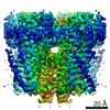 21586MC M: map data used to model this data C: citing same article ( |
|---|---|
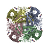
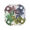
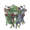
| Similar structure data |


-Links
 PDBj
PDBj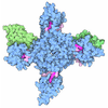
- Assembly
| Deposited unit | 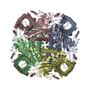
|
|---|---|
| 1 |
|
-Components
| #1: Protein | Mass: 86517.031 Da / Num. of mol.: 4 / Mutation: C331S Source method: isolated from a genetically manipulated source Source: (gene. exp.) Homo sapiens (human) / Gene: PKD2, TRPP2 / Production host: Homo sapiens (human) / Strain (production host): HEK293S GnTi- / References: UniProt: Q13563#2: Sugar | ChemComp-NAG /   Type: D-saccharide, beta linking / Mass: 221.208 Da / Num. of mol.: 12 Type: D-saccharide, beta linking / Mass: 221.208 Da / Num. of mol.: 12Source method: isolated from a genetically manipulated source Formula: C8H15NO6 / Feature type: SUBJECT OF INVESTIGATION Has ligand of interest | Y | Has protein modification | Y | |
|---|
-Experimental details
-Experiment
| Experiment | Method: ELECTRON MICROSCOPY |
|---|---|
| EM experiment | Aggregation state: PARTICLE / 3D reconstruction method: single particle reconstruction |
- Sample preparation
| Component | Name: PKD2 / Type: CELL / Entity ID: #1 / Source: RECOMBINANT |
|---|---|
| Source (natural) | Organism: Homo sapiens (human) |
| Source (recombinant) | Organism: Homo sapiens (human) / Strain: HEK293S |
| Buffer solution | pH: 7.4 |
| Specimen | Conc.: 2 mg/ml / Embedding applied: NO / Shadowing applied: NO / Staining applied: NO / Vitrification applied: YES |
| Vitrification | Cryogen name: ETHANE |
- Electron microscopy imaging
| Experimental equipment |  Model: Titan Krios / Image courtesy: FEI Company |
|---|---|
| Microscopy | Model: FEI TITAN KRIOS |
| Electron gun | Electron source:  FIELD EMISSION GUN / Accelerating voltage: 300 kV / Illumination mode: FLOOD BEAM FIELD EMISSION GUN / Accelerating voltage: 300 kV / Illumination mode: FLOOD BEAM |
| Electron lens | Mode: DARK FIELD |
| Image recording | Electron dose: 1 e/Å2 / Film or detector model: GATAN K2 IS (4k x 4k) |
- Processing
| Software | Name: PHENIX / Version: 1.10.1_2155: / Classification: refinement | ||||||||||||||||||||||||
|---|---|---|---|---|---|---|---|---|---|---|---|---|---|---|---|---|---|---|---|---|---|---|---|---|---|
| EM software | Name: PHENIX / Category: model refinement | ||||||||||||||||||||||||
| CTF correction | Type: PHASE FLIPPING AND AMPLITUDE CORRECTION | ||||||||||||||||||||||||
| 3D reconstruction | Resolution: 3.24 Å / Resolution method: FSC 0.143 CUT-OFF / Num. of particles: 74302 / Symmetry type: POINT | ||||||||||||||||||||||||
| Atomic model building | B value: 100 / Protocol: OTHER / Space: REAL | ||||||||||||||||||||||||
| Refine LS restraints |
|
