 Movie
Movie Controller


+ Open data

- Basic information
| Entry | Database: PDB / ID: 6vqq | ||||||
|---|---|---|---|---|---|---|---|
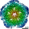
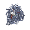
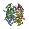
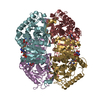
| Title | CryoEM Structure of the Plasmodium falciparum transporter PfFNT | ||||||
 Components Components | Formate-nitrite transporter | ||||||
 Keywords Keywords | MEMBRANE PROTEIN / Transporter | ||||||
| Function / homology |  Function and homology information Function and homology informationhigh-affinity secondary active nitrite transmembrane transporter activity / lactate transmembrane transport / nitrite transport / lactate:proton symporter activity / vacuolar membrane / plasma membrane Similarity search - Function | ||||||
| Biological species |  | ||||||
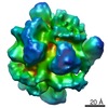
| Method | ELECTRON MICROSCOPY / single particle reconstruction / cryo EM / Resolution: 2.56 Å | ||||||
 Authors Authors | Su, C.C. / Lyu, M. | ||||||
 Citation Citation | Journal: EMBO Rep / Year: 2021 Title: Structural basis of transport and inhibition of the Plasmodium falciparum transporter PfFNT. Authors: Meinan Lyu / Chih-Chia Su / James W Kazura / Edward W Yu /  Abstract: The intra-erythrocyte stage of P. falciparum relies primarily on glycolysis to generate adenosine triphosphate (ATP) and the energy required to support growth and reproduction. Lactic acid, a ...The intra-erythrocyte stage of P. falciparum relies primarily on glycolysis to generate adenosine triphosphate (ATP) and the energy required to support growth and reproduction. Lactic acid, a metabolic byproduct of glycolysis, is potentially toxic as it lowers the pH inside the parasite. Plasmodium falciparum formate-nitrite transporter (PfFNT), a 34-kDa transmembrane protein, has been identified as a novel drug target as it exports lactate from inside the parasite to the surrounding parasitophorous vacuole within the erythrocyte cytosol. The structure and detailed molecular mechanism of this membrane protein are not yet available. Here we present structures of PfFNT in the absence and presence of the functional inhibitor MMV007839 at resolutions of 2.56 Å and 2.78 Å using single-particle cryo-electron microscopy. Genetic analysis and transport assay indicate that PfFNT is able to transfer lactate across the membrane. Combined, our data suggest a stepwise displacement mechanism for substrate transport. The PfFNT membrane protein is capable of picking up lactate ions from the parasite's cytosol, converting them to lactic acids and then exporting these acids into the extracellular space. | ||||||
| History |
|
- Structure visualization
| Movie |
Movie viewer |
|---|---|
| Structure viewer | Molecule: MolmilJmol/JSmol |
- Downloads & links
-Download
| PDBx/mmCIF format | 6vqq.cif.gz | 236.9 KB | Display | PDBx/mmCIF format |
|---|---|---|---|---|
| PDB format | pdb6vqq.ent.gz | 196.3 KB | Display | PDB format |
| PDBx/mmJSON format | 6vqq.json.gz | Tree view | PDBx/mmJSON format | |
| Others |  Other downloads Other downloads |
-Validation report
| Arichive directory | https://data.pdbj.org/pub/pdb/validation_reports/vq/6vqqftp://data.pdbj.org/pub/pdb/validation_reports/vq/6vqq | HTTPS FTP |
|---|
-Related structure data
| Related structure data | 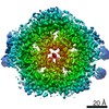 21354MC 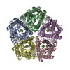 6vqrC 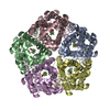 7mxyC M: map data used to model this data C: citing same article ( |
|---|---|
| Similar structure data |


-Links
 PDBj
PDBj
- Assembly
| Deposited unit | 
|
|---|---|
| 1 |
|
-Components
| #1: Protein | Mass: 34492.281 Da / Num. of mol.: 5 Source method: isolated from a genetically manipulated source Source: (gene. exp.) Strain: isolate 3D7 / Gene: PF3D7_0316600 / Production host:  Homo sapiens (human) / References: UniProt: O77389 Homo sapiens (human) / References: UniProt: O77389 |
|---|
-Experimental details
-Experiment
| Experiment | Method: ELECTRON MICROSCOPY |
|---|---|
| EM experiment | Aggregation state: PARTICLE / 3D reconstruction method: single particle reconstruction |
- Sample preparation
| Component | Name: PfFNT / Type: COMPLEX / Entity ID: all / Source: RECOMBINANT |
|---|---|
| Source (natural) | Organism: |
| Source (recombinant) | Organism: Homo sapiens (human) |
| Buffer solution | pH: 7.5 |
| Specimen | Conc.: 0.5 mg/ml / Embedding applied: NO / Shadowing applied: NO / Staining applied: NO / Vitrification applied: YES / Details: This sample was monodisperse. |
| Specimen support | Grid material: COPPER / Grid mesh size: 400 divisions/in. / Grid type: Quantifoil R1.2/1.3 |
| Vitrification | Instrument: FEI VITROBOT MARK IV / Cryogen name: ETHANE / Humidity: 100 % / Chamber temperature: 277 K |
- Electron microscopy imaging
| Experimental equipment |  Model: Titan Krios / Image courtesy: FEI Company |
|---|---|
| Microscopy | Model: FEI TITAN KRIOS |
| Electron gun | Electron source:  FIELD EMISSION GUN / Accelerating voltage: 300 kV / Illumination mode: SPOT SCAN FIELD EMISSION GUN / Accelerating voltage: 300 kV / Illumination mode: SPOT SCAN |
| Electron lens | Mode: BRIGHT FIELD |
| Image recording | Electron dose: 29 e/Å2 / Film or detector model: GATAN K3 BIOQUANTUM (6k x 4k) |
- Processing
| CTF correction | Type: PHASE FLIPPING AND AMPLITUDE CORRECTION |
|---|---|
| 3D reconstruction | Resolution: 2.56 Å / Resolution method: FSC 0.143 CUT-OFF / Num. of particles: 85976 / Symmetry type: POINT |
