 Movie
Movie Controller


+ Open data

- Basic information
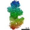
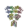
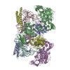
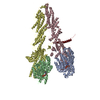
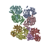
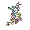
| Entry | Database: PDB / ID: 6um2 | ||||||
|---|---|---|---|---|---|---|---|
| Title | Structure of M-6-P/IGFII Receptor and IGFII complex | ||||||
 Components Components |
| ||||||
 Keywords Keywords | SUGAR BINDING PROTEIN / M-6-P / IGFII / receptor | ||||||
| Function / homology |  Function and homology information Function and homology informationkringle domain binding / embryonic placenta morphogenesis / negative regulation of muscle cell differentiation / positive regulation of skeletal muscle tissue growth / insulin-like growth factor binding / Signaling by Type 1 Insulin-like Growth Factor 1 Receptor (IGF1R) / regulation of muscle cell differentiation / IRS-related events triggered by IGF1R / positive regulation of organ growth / transmembrane receptor protein tyrosine kinase activator activity ...kringle domain binding / embryonic placenta morphogenesis / negative regulation of muscle cell differentiation / positive regulation of skeletal muscle tissue growth / insulin-like growth factor binding / Signaling by Type 1 Insulin-like Growth Factor 1 Receptor (IGF1R) / regulation of muscle cell differentiation / IRS-related events triggered by IGF1R / positive regulation of organ growth / transmembrane receptor protein tyrosine kinase activator activity / genomic imprinting / positive regulation of multicellular organism growth / exocrine pancreas development / positive regulation of activated T cell proliferation / positive regulation of vascular endothelial cell proliferation / lysosomal transport / D-mannose binding / positive regulation of cell division / positive regulation of glycogen biosynthetic process / positive regulation of insulin receptor signaling pathway / embryonic placenta development / endocytic vesicle / SHC-related events triggered by IGF1R / insulin-like growth factor receptor binding / striated muscle cell differentiation / positive regulation of mitotic nuclear division / insulin-like growth factor receptor signaling pathway / platelet alpha granule lumen / animal organ morphogenesis / insulin receptor binding / protein serine/threonine kinase activator activity / growth factor activity / trans-Golgi network / phosphoprotein binding / hormone activity / integrin binding / Regulation of Insulin-like Growth Factor (IGF) transport and uptake by Insulin-like Growth Factor Binding Proteins (IGFBPs) / glucose metabolic process / osteoblast differentiation / insulin receptor signaling pathway / late endosome / Platelet degranulation / signaling receptor activity / in utero embryonic development / positive regulation of MAPK cascade / positive regulation of phosphatidylinositol 3-kinase/protein kinase B signal transduction / endosome membrane / receptor ligand activity / Golgi membrane / positive regulation of cell population proliferation / regulation of DNA-templated transcription / cell surface / negative regulation of transcription by RNA polymerase II / Golgi apparatus / positive regulation of transcription by RNA polymerase II / : / extracellular region / plasma membrane Similarity search - Function | ||||||
| Biological species |  Homo sapiens (human) Homo sapiens (human) | ||||||
| Method | ELECTRON MICROSCOPY / single particle reconstruction / cryo EM / Resolution: 4.32 Å | ||||||
 Authors Authors | Wang, R. / Qi, X. / Li, X. | ||||||
 Citation Citation | Journal: Sci Adv / Year: 2020 Title: Marked structural rearrangement of mannose 6-phosphate/IGF2 receptor at different pH environments. Authors: Rong Wang / Xiaofeng Qi / Philip Schmiege / Elias Coutavas / Xiaochun Li /  Abstract: Many cell surface receptors internalize their ligands and deliver them to endosomes, where the acidic pH causes the ligand to dissociate. The liberated receptor returns to the cell surface in a ...Many cell surface receptors internalize their ligands and deliver them to endosomes, where the acidic pH causes the ligand to dissociate. The liberated receptor returns to the cell surface in a process called receptor cycling. The structural basis for pH-dependent ligand dissociation is not well understood. In some receptors, the ligand binding domain is composed of multiple repeated sequences. The insulin-like growth factor 2 receptor (IGF2R) contains 15 β strand-rich repeat domains. The overall structure and the mechanism by which IGF2R binds IGF2 and releases it are unknown. We used cryo-EM to determine the structures of the IGF2R at pH 7.4 with IGF2 bound and at pH 4.5 in the ligand-dissociated state. The results reveal different arrangements of the receptor in different pH environments mediated by changes in the interactions between the repeated sequences. These results have implications for our understanding of ligand release from receptors in endocytic compartments. | ||||||
| History |
|
- Structure visualization
| Movie |
Movie viewer |
|---|---|
| Structure viewer | Molecule: MolmilJmol/JSmol |
- Downloads & links
-Download
| PDBx/mmCIF format | 6um2.cif.gz | 333.3 KB | Display | PDBx/mmCIF format |
|---|---|---|---|---|
| PDB format | pdb6um2.ent.gz | 249.6 KB | Display | PDB format |
| PDBx/mmJSON format | 6um2.json.gz | Tree view | PDBx/mmJSON format | |
| Others |  Other downloads Other downloads |
-Validation report
| Arichive directory | https://data.pdbj.org/pub/pdb/validation_reports/um/6um2ftp://data.pdbj.org/pub/pdb/validation_reports/um/6um2 | HTTPS FTP |
|---|
-Related structure data
| Related structure data |  20816MC 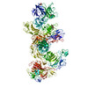 6um1C M: map data used to model this data C: citing same article ( |
|---|---|
| Similar structure data |
-Links
 PDBj
PDBj


- Assembly
| Deposited unit | 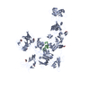
|
|---|---|
| 1 |
|
-Components
| #1: Protein | Mass: 274830.125 Da / Num. of mol.: 1 / Source method: isolated from a natural source / Source: (natural) | ||||
|---|---|---|---|---|---|
| #2: Protein | Mass: 7615.667 Da / Num. of mol.: 1 Source method: isolated from a genetically manipulated source Source: (gene. exp.) Homo sapiens (human) / Gene: IGF2, PP1446 / Production host:  | ||||
| #3: Sugar | ChemComp-NAG /   Type: D-saccharide, beta linking / Mass: 221.208 Da / Num. of mol.: 7 Type: D-saccharide, beta linking / Mass: 221.208 Da / Num. of mol.: 7Source method: isolated from a genetically manipulated source Formula: C8H15NO6 Has ligand of interest | N | Has protein modification | Y | |
-Experimental details
-Experiment
| Experiment | Method: ELECTRON MICROSCOPY |
|---|---|
| EM experiment | Aggregation state: PARTICLE / 3D reconstruction method: single particle reconstruction |
- Sample preparation
| Component |
| ||||||||||||||||||||||||
|---|---|---|---|---|---|---|---|---|---|---|---|---|---|---|---|---|---|---|---|---|---|---|---|---|---|
| Source (natural) |
| ||||||||||||||||||||||||
| Source (recombinant) | Organism: | ||||||||||||||||||||||||
| Buffer solution | pH: 7.4 | ||||||||||||||||||||||||
| Specimen | Embedding applied: NO / Shadowing applied: NO / Staining applied: NO / Vitrification applied: YES | ||||||||||||||||||||||||
| Vitrification | Cryogen name: ETHANE |
- Electron microscopy imaging
| Experimental equipment |  Model: Titan Krios / Image courtesy: FEI Company |
|---|---|
| Microscopy | Model: FEI TITAN KRIOS |
| Electron gun | Electron source:  FIELD EMISSION GUN / Accelerating voltage: 300 kV / Illumination mode: FLOOD BEAM FIELD EMISSION GUN / Accelerating voltage: 300 kV / Illumination mode: FLOOD BEAM |
| Electron lens | Mode: DARK FIELD |
| Image recording | Electron dose: 100 e/Å2 / Film or detector model: GATAN K3 (6k x 4k) |
- Processing
| Software | Name: REFMAC / Version: 5.8.0238 / Classification: refinement | ||||||||||||||||||||||||||||||||||||||||||||||||||||||||||||||||||||||||||||||||||||||||||||||||||||||||||
|---|---|---|---|---|---|---|---|---|---|---|---|---|---|---|---|---|---|---|---|---|---|---|---|---|---|---|---|---|---|---|---|---|---|---|---|---|---|---|---|---|---|---|---|---|---|---|---|---|---|---|---|---|---|---|---|---|---|---|---|---|---|---|---|---|---|---|---|---|---|---|---|---|---|---|---|---|---|---|---|---|---|---|---|---|---|---|---|---|---|---|---|---|---|---|---|---|---|---|---|---|---|---|---|---|---|---|---|
| CTF correction | Type: NONE | ||||||||||||||||||||||||||||||||||||||||||||||||||||||||||||||||||||||||||||||||||||||||||||||||||||||||||
| 3D reconstruction | Resolution: 4.32 Å / Resolution method: FSC 0.143 CUT-OFF / Num. of particles: 75821 / Symmetry type: POINT | ||||||||||||||||||||||||||||||||||||||||||||||||||||||||||||||||||||||||||||||||||||||||||||||||||||||||||
| Refinement | Resolution: 4.32→302.72 Å / Cor.coef. Fo:Fc: 0.76 / SU B: 65.401 / SU ML: 0.813 / ESU R: 0.29 / Stereochemistry target values: MAXIMUM LIKELIHOOD / Details: HYDROGENS HAVE BEEN ADDED IN THE RIDING POSITIONS
| ||||||||||||||||||||||||||||||||||||||||||||||||||||||||||||||||||||||||||||||||||||||||||||||||||||||||||
| Solvent computation | Ion probe radii: 0.8 Å / Shrinkage radii: 0.8 Å / VDW probe radii: 1.2 Å / Solvent model: MASK | ||||||||||||||||||||||||||||||||||||||||||||||||||||||||||||||||||||||||||||||||||||||||||||||||||||||||||
| Displacement parameters | Biso mean: 19.059 Å2
| ||||||||||||||||||||||||||||||||||||||||||||||||||||||||||||||||||||||||||||||||||||||||||||||||||||||||||
| Refinement step | Cycle: 1 / Total: 13094 | ||||||||||||||||||||||||||||||||||||||||||||||||||||||||||||||||||||||||||||||||||||||||||||||||||||||||||
| Refine LS restraints |
|