 Movie
Movie Controller


+ Open data

- Basic information
| Entry | Database: PDB / ID: 6ukp | |||||||||||||||||||||||||||||||||
|---|---|---|---|---|---|---|---|---|---|---|---|---|---|---|---|---|---|---|---|---|---|---|---|---|---|---|---|---|---|---|---|---|---|---|
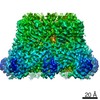
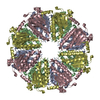
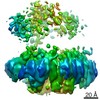
| Title | Apo mBcs1 | |||||||||||||||||||||||||||||||||
 Components Components | Mitochondrial chaperone BCS1 | |||||||||||||||||||||||||||||||||
 Keywords Keywords | CHAPERONE / bcs1 / AAA ATPases / mitochondrial inner membrane | |||||||||||||||||||||||||||||||||
| Function / homology |  Function and homology information Function and homology informationmitochondrial protein-transporting ATPase activity / protein insertion into mitochondrial inner membrane from matrix / mitochondrial respiratory chain complex IV assembly / mitochondrial respiratory chain complex III assembly / mitochondrial respiratory chain complex I assembly / Hydrolases; Acting on acid anhydrides; In phosphorus-containing anhydrides / mitochondrion organization / mitochondrial inner membrane / ATP hydrolysis activity / mitochondrion / ATP binding Similarity search - Function | |||||||||||||||||||||||||||||||||
| Biological species |  | |||||||||||||||||||||||||||||||||
| Method | ELECTRON MICROSCOPY / single particle reconstruction / cryo EM / Resolution: 3.81 Å | |||||||||||||||||||||||||||||||||
 Authors Authors | Tang, W.K. / Borgnia, M.J. / Hsu, A.L. / Xia, D. | |||||||||||||||||||||||||||||||||
| Funding support |  United States, 1items United States, 1items
| |||||||||||||||||||||||||||||||||
 Citation Citation | Journal: Nat Struct Mol Biol / Year: 2020 Title: Structures of AAA protein translocase Bcs1 suggest translocation mechanism of a folded protein. Authors: Wai Kwan Tang / Mario J Borgnia / Allen L Hsu / Lothar Esser / Tara Fox / Natalia de Val / Di Xia / Abstract: The mitochondrial membrane-bound AAA protein Bcs1 translocate substrates across the mitochondrial inner membrane without previous unfolding. One substrate of Bcs1 is the iron-sulfur protein (ISP), a ...The mitochondrial membrane-bound AAA protein Bcs1 translocate substrates across the mitochondrial inner membrane without previous unfolding. One substrate of Bcs1 is the iron-sulfur protein (ISP), a subunit of the respiratory Complex III. How Bcs1 translocates ISP across the membrane is unknown. Here we report structures of mouse Bcs1 in two different conformations, representing three nucleotide states. The apo and ADP-bound structures reveal a homo-heptamer and show a large putative substrate-binding cavity accessible to the matrix space. ATP binding drives a contraction of the cavity by concerted motion of the ATPase domains, which could push substrate across the membrane. Our findings shed light on the potential mechanism of translocating folded proteins across a membrane, offer insights into the assembly process of Complex III and allow mapping of human disease-associated mutations onto the Bcs1 structure. | |||||||||||||||||||||||||||||||||
| History |
|
- Structure visualization
| Movie |
Movie viewer |
|---|---|
| Structure viewer | Molecule: MolmilJmol/JSmol |
- Downloads & links
-Download
| PDBx/mmCIF format | 6ukp.cif.gz | 834.2 KB | Display | PDBx/mmCIF format |
|---|---|---|---|---|
| PDB format | pdb6ukp.ent.gz | 705.5 KB | Display | PDB format |
| PDBx/mmJSON format | 6ukp.json.gz | Tree view | PDBx/mmJSON format | |
| Others |  Other downloads Other downloads |
-Validation report
| Arichive directory | https://data.pdbj.org/pub/pdb/validation_reports/uk/6ukpftp://data.pdbj.org/pub/pdb/validation_reports/uk/6ukp | HTTPS FTP |
|---|
-Related structure data
| Related structure data | 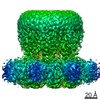 20808MC 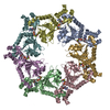 6u1yC 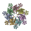 6ukoC 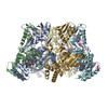 6uksC M: map data used to model this data C: citing same article ( |
|---|---|
| Similar structure data |


-Links
 PDBj
PDBj


- Assembly
| Deposited unit | 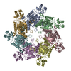
|
|---|---|
| 1 |
|
-Components
| #1: Protein | Mass: 48659.281 Da / Num. of mol.: 7 Source method: isolated from a genetically manipulated source Source: (gene. exp.)  Komagataella pastoris (fungus) / References: UniProt: Q9CZP5 Komagataella pastoris (fungus) / References: UniProt: Q9CZP5Has protein modification | N | |
|---|
-Experimental details
-Experiment
| Experiment | Method: ELECTRON MICROSCOPY |
|---|---|
| EM experiment | Aggregation state: PARTICLE / 3D reconstruction method: single particle reconstruction |
- Sample preparation
| Component | Name: Apo structure of mBcs1 heptamer / Type: COMPLEX / Entity ID: all / Source: RECOMBINANT |
|---|---|
| Source (natural) | Organism: |
| Source (recombinant) | Organism: Komagataella pastoris (fungus) |
| Buffer solution | pH: 8.5 |
| Specimen | Embedding applied: NO / Shadowing applied: NO / Staining applied: NO / Vitrification applied: YES |
| Vitrification | Cryogen name: ETHANE |
- Electron microscopy imaging
| Experimental equipment |  Model: Titan Krios / Image courtesy: FEI Company |
|---|---|
| Microscopy | Model: FEI TITAN KRIOS |
| Electron gun | Electron source:  FIELD EMISSION GUN / Accelerating voltage: 300 kV / Illumination mode: FLOOD BEAM FIELD EMISSION GUN / Accelerating voltage: 300 kV / Illumination mode: FLOOD BEAM |
| Electron lens | Mode: BRIGHT FIELD |
| Image recording | Electron dose: 40 e/Å2 / Film or detector model: GATAN K2 SUMMIT (4k x 4k) |
- Processing
| Software | Name: PHENIX / Version: 1.16_3549: / Classification: refinement | ||||||||||||||||||||||||
|---|---|---|---|---|---|---|---|---|---|---|---|---|---|---|---|---|---|---|---|---|---|---|---|---|---|
| EM software | Name: PHENIX / Category: model refinement | ||||||||||||||||||||||||
| CTF correction | Type: PHASE FLIPPING AND AMPLITUDE CORRECTION | ||||||||||||||||||||||||
| 3D reconstruction | Resolution: 3.81 Å / Resolution method: FSC 0.143 CUT-OFF / Num. of particles: 67261 / Symmetry type: POINT | ||||||||||||||||||||||||
| Refine LS restraints |
|
