 ムービー
ムービー コントローラー


+ データを開く

- 基本情報
基本情報
| 登録情報 | データベース: PDB / ID: 6u5o | ||||||||||||||||||||||||||||||||||||||||||||||||||||||
|---|---|---|---|---|---|---|---|---|---|---|---|---|---|---|---|---|---|---|---|---|---|---|---|---|---|---|---|---|---|---|---|---|---|---|---|---|---|---|---|---|---|---|---|---|---|---|---|---|---|---|---|---|---|---|---|
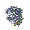
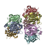
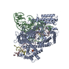
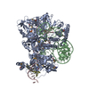
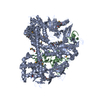
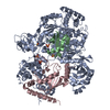
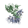
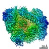
| タイトル | Structure of the Human Metapneumovirus Polymerase bound to the phosphoprotein tetramer | ||||||||||||||||||||||||||||||||||||||||||||||||||||||
 要素 要素 |
| ||||||||||||||||||||||||||||||||||||||||||||||||||||||
 キーワード キーワード | VIRAL PROTEIN / human metapneumovirus / HMPV / Polymerase / Phosphoprotein / Respiratory syncytial virus / RSV / pneumoviridae / Mononegavirales | ||||||||||||||||||||||||||||||||||||||||||||||||||||||
| 機能・相同性 |  機能・相同性情報 機能・相同性情報NNS virus cap methyltransferase / GDP polyribonucleotidyltransferase / 加水分解酵素; 酸無水物に作用; リン含有酸無水物に作用 / virion component / mRNA 5'-cap (guanine-N7-)-methyltransferase activity / host cell cytoplasm / RNA-directed RNA polymerase / RNA-directed RNA polymerase activity / GTPase activity / ATP binding / metal ion binding 類似検索 - 分子機能 | ||||||||||||||||||||||||||||||||||||||||||||||||||||||
| 生物種 |  Human metapneumovirus (ウイルス) Human metapneumovirus (ウイルス) | ||||||||||||||||||||||||||||||||||||||||||||||||||||||
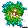
| 手法 | 電子顕微鏡法 / 単粒子再構成法 / クライオ電子顕微鏡法 / 解像度: 3.7 Å | ||||||||||||||||||||||||||||||||||||||||||||||||||||||
 データ登録者 データ登録者 | Pan, J. / Qian, X. / Lattmann, S. / Sahili, A.E. / Yeo, T.H. / Kalocsay, M. / Fearns, R. / Lescar, J. | ||||||||||||||||||||||||||||||||||||||||||||||||||||||
| 資金援助 |  シンガポール, シンガポール,  米国, 3件 米国, 3件
| ||||||||||||||||||||||||||||||||||||||||||||||||||||||
 引用 引用 | ジャーナル: Nature / 年: 2020 タイトル: Structure of the human metapneumovirus polymerase phosphoprotein complex. 著者: Junhua Pan / Xinlei Qian / Simon Lattmann / Abbas El Sahili / Tiong Han Yeo / Huan Jia / Tessa Cressey / Barbara Ludeke / Sarah Noton / Marian Kalocsay / Rachel Fearns / Julien Lescar / 要旨: Respiratory syncytial virus (RSV) and human metapneumovirus (HMPV) cause severe respiratory diseases in infants and elderly adults. No vaccine or effective antiviral therapy currently exists to ...Respiratory syncytial virus (RSV) and human metapneumovirus (HMPV) cause severe respiratory diseases in infants and elderly adults. No vaccine or effective antiviral therapy currently exists to control RSV or HMPV infections. During viral genome replication and transcription, the tetrameric phosphoprotein P serves as a crucial adaptor between the ribonucleoprotein template and the L protein, which has RNA-dependent RNA polymerase (RdRp), GDP polyribonucleotidyltransferase and cap-specific methyltransferase activities. How P interacts with L and mediates the association with the free form of N and with the ribonucleoprotein is not clear for HMPV or other major human pathogens, including the viruses that cause measles, Ebola and rabies. Here we report a cryo-electron microscopy reconstruction that shows the ring-shaped structure of the polymerase and capping domains of HMPV-L bound to a tetramer of P. The connector and methyltransferase domains of L are mobile with respect to the core. The putative priming loop that is important for the initiation of RNA synthesis is fully retracted, which leaves space in the active-site cavity for RNA elongation. P interacts extensively with the N-terminal region of L, burying more than 4,016 Å of the molecular surface area in the interface. Two of the four helices that form the coiled-coil tetramerization domain of P, and long C-terminal extensions projecting from these two helices, wrap around the L protein in a manner similar to tentacles. The structural versatility of the four P protomers-which are largely disordered in their free state-demonstrates an example of a 'folding-upon-partner-binding' mechanism for carrying out P adaptor functions. The structure shows that P has the potential to modulate multiple functions of L and these results should accelerate the design of specific antiviral drugs. | ||||||||||||||||||||||||||||||||||||||||||||||||||||||
| 履歴 |
|
- 構造の表示
構造の表示
| ムービー |
ムービービューア |
|---|---|
| 構造ビューア | 分子: MolmilJmol/JSmol |
- ダウンロードとリンク
-ダウンロード
| PDBx/mmCIF形式 | 6u5o.cif.gz | 579.9 KB | 表示 | PDBx/mmCIF形式 |
|---|---|---|---|---|
| PDB形式 | pdb6u5o.ent.gz | 458.9 KB | 表示 | PDB形式 |
| PDBx/mmJSON形式 | 6u5o.json.gz | ツリー表示 | PDBx/mmJSON形式 | |
| その他 |  その他のダウンロード その他のダウンロード |
-検証レポート
| アーカイブディレクトリ | https://data.pdbj.org/pub/pdb/validation_reports/u5/6u5oftp://data.pdbj.org/pub/pdb/validation_reports/u5/6u5o | HTTPS FTP |
|---|
-関連構造データ


-リンク
 PDBj
PDBj
- 集合体
集合体
| 登録構造単位 | 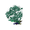
|
|---|---|
| 1 |
|
-要素
| #1: タンパク質 | 分子量: 233889.562 Da / 分子数: 1 / 由来タイプ: 組換発現 詳細: amino acid residues 1-25 is the N-terminal expression/purification tag containing a Strep II tag, a linker and a TEV protease cleavage site. 由来: (組換発現) Human metapneumovirus (strain CAN97-83) (ウイルス)株: CAN97-83 / 細胞株 (発現宿主): Sf9 発現宿主:   Spodoptera frugiperda (ツマジロクサヨトウ) Spodoptera frugiperda (ツマジロクサヨトウ)株 (発現宿主): IPLB-Sf21-AE 参照: UniProt: Q6WB93, RNA-directed RNA polymerase, mRNA (guanine-N7)-methyltransferase, 転移酵素; リンを含む基を移すもの; 核酸を移すもの, GDP polyribonucleotidyltransferase | ||
|---|---|---|---|
| #2: タンパク質 | 分子量: 35685.066 Da / 分子数: 4 / 由来タイプ: 組換発現 由来: (組換発現) Human metapneumovirus (strain CAN97-83) (ウイルス)株: CAN97-83 / 細胞株 (発現宿主): Sf9 発現宿主: Spodoptera frugiperda (ツマジロクサヨトウ)株 (発現宿主): IPLB-Sf21-AE / 参照: UniProt: Q8B9Q8 Has protein modification | N | |
-実験情報
-実験
| 実験 | 手法: 電子顕微鏡法 |
|---|---|
| EM実験 | 試料の集合状態: PARTICLE / 3次元再構成法: 単粒子再構成法 |
- 試料調製
試料調製
| 構成要素 | 名称: Human metapneumovirus CAN97-83 / タイプ: COMPLEX 詳細: Human metapneumovirus L and P genes were co-expressed in sf9 insect cells using recombinant baculovirus expression system. The protein complex were affinity purified using nickel-NTA resin, ...詳細: Human metapneumovirus L and P genes were co-expressed in sf9 insect cells using recombinant baculovirus expression system. The protein complex were affinity purified using nickel-NTA resin, followed by purification on a HiTrap Heparin column and a superpose 6 size exclusion column. Entity ID: all / 由来: RECOMBINANT | |||||||||||||||||||||||||
|---|---|---|---|---|---|---|---|---|---|---|---|---|---|---|---|---|---|---|---|---|---|---|---|---|---|---|
| 分子量 | 値: 0.36 MDa / 実験値: YES | |||||||||||||||||||||||||
| 由来(天然) | 生物種: Human metapneumovirus CAN97-83 (ウイルス) | |||||||||||||||||||||||||
| 由来(組換発現) | 生物種: Spodoptera frugiperda (ツマジロクサヨトウ) 株: IPLB-Sf21-AE / 細胞: sf9 / プラスミド: pFastBacDual | |||||||||||||||||||||||||
| 天然宿主 | 生物種: homo sa | |||||||||||||||||||||||||
| 緩衝液 | pH: 7.4 詳細: sample of protein complex was in buffer containing 20 mM Na Hepes, pH 7.4, 300 mM NaCl, 1 mM MgCl2 and 0.5 mM TCEP (components as listed). | |||||||||||||||||||||||||
| 緩衝液成分 |
| |||||||||||||||||||||||||
| 試料 | 濃度: 0.8 mg/ml / 包埋: NO / シャドウイング: NO / 染色: NO / 凍結: YES 詳細: purified human metapneumovirus L:P complex in a 1:4 stoichiometry. | |||||||||||||||||||||||||
| 試料支持 | 詳細: 20 mA / グリッドの材料: COPPER / グリッドのサイズ: 200 divisions/in. / グリッドのタイプ: Quantifoil R1.2/1.3 | |||||||||||||||||||||||||
| 急速凍結 | 装置: FEI VITROBOT MARK IV / 凍結剤: ETHANE / 湿度: 100 % / 凍結前の試料温度: 298 K 詳細: 3.5 uL sample (0.8 mg/mL)/grid, 4-second blotting (double-sided) at offset -2 before plunging. |
- 電子顕微鏡撮影
| 実験機器 |  モデル: Tecnai Polara / 画像提供: FEI Company |
|---|---|
| 顕微鏡 | モデル: FEI POLARA 300 詳細: preliminary grid screening was performed manually on a FEI Tecnai F20 microscope equipped with a Gatan K2 summit camera. |
| 電子銃 | 電子線源:  FIELD EMISSION GUN / 加速電圧: 300 kV / 照射モード: OTHER FIELD EMISSION GUN / 加速電圧: 300 kV / 照射モード: OTHER |
| 電子レンズ | モード: BRIGHT FIELD / 倍率(公称値): 31000 X / 倍率(補正後): 40323 X / 最大 デフォーカス(公称値): 3000 nm / 最小 デフォーカス(公称値): 1500 nm / Calibrated defocus min: 1500 nm / 最大 デフォーカス(補正後): 3000 nm / Cs: 2.26 mm / C2レンズ絞り径: 50 µm / アライメント法: COMA FREE |
| 試料ホルダ | 凍結剤: NITROGEN / 試料ホルダーモデル: OTHER / 最高温度: 90 K / 最低温度: 77 K |
| 撮影 | 平均露光時間: 0.2 sec. / 電子線照射量: 1.2 e/Å2 / 検出モード: SUPER-RESOLUTION フィルム・検出器のモデル: GATAN K2 SUMMIT (4k x 4k) 撮影したグリッド数: 1 / 実像数: 7438 |
| 画像スキャン | サンプリングサイズ: 5 µm / 横: 7676 / 縦: 7420 / 動画フレーム数/画像: 40 / 利用したフレーム数/画像: 1-40 |
- 解析
解析
| ソフトウェア | 名称: PHENIX / バージョン: 1.16_3549: / 分類: 精密化 | ||||||||||||||||||||||||||||||||||||||||
|---|---|---|---|---|---|---|---|---|---|---|---|---|---|---|---|---|---|---|---|---|---|---|---|---|---|---|---|---|---|---|---|---|---|---|---|---|---|---|---|---|---|
| EMソフトウェア |
| ||||||||||||||||||||||||||||||||||||||||
| CTF補正 | タイプ: NONE | ||||||||||||||||||||||||||||||||||||||||
| 粒子像の選択 | 選択した粒子像数: 5381943 | ||||||||||||||||||||||||||||||||||||||||
| 対称性 | 点対称性: C1 (非対称) | ||||||||||||||||||||||||||||||||||||||||
| 3次元再構成 | 解像度: 3.7 Å / 解像度の算出法: FSC 0.143 CUT-OFF / 粒子像の数: 302346 / 対称性のタイプ: POINT | ||||||||||||||||||||||||||||||||||||||||
| 拘束条件 |
|
