 Movie
Movie Controller

[English] 日本語
 Yorodumi
- PDB-6u5o: Structure of the Human Metapneumovirus Polymerase bound to the ph... -
+ Open data

- Basic information
| Entry | Database: PDB / ID: 6u5o | ||||||||||||||||||||||||||||||||||||||||||||||||||||||
|---|---|---|---|---|---|---|---|---|---|---|---|---|---|---|---|---|---|---|---|---|---|---|---|---|---|---|---|---|---|---|---|---|---|---|---|---|---|---|---|---|---|---|---|---|---|---|---|---|---|---|---|---|---|---|---|
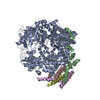
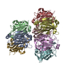
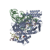
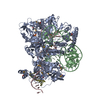
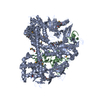
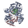
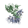
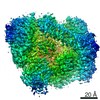
| Title | Structure of the Human Metapneumovirus Polymerase bound to the phosphoprotein tetramer | ||||||||||||||||||||||||||||||||||||||||||||||||||||||
 Components Components |
| ||||||||||||||||||||||||||||||||||||||||||||||||||||||
 Keywords Keywords | VIRAL PROTEIN / human metapneumovirus / HMPV / Polymerase / Phosphoprotein / Respiratory syncytial virus / RSV / pneumoviridae / Mononegavirales | ||||||||||||||||||||||||||||||||||||||||||||||||||||||
| Function / homology |  Function and homology information Function and homology informationNNS virus cap methyltransferase / GDP polyribonucleotidyltransferase / Hydrolases; Acting on acid anhydrides; In phosphorus-containing anhydrides / virion component / mRNA 5'-cap (guanine-N7-)-methyltransferase activity / host cell cytoplasm / RNA-directed RNA polymerase / RNA-directed RNA polymerase activity / GTPase activity / ATP binding / metal ion binding Similarity search - Function | ||||||||||||||||||||||||||||||||||||||||||||||||||||||
| Biological species |  Human metapneumovirus Human metapneumovirus | ||||||||||||||||||||||||||||||||||||||||||||||||||||||
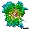
| Method | ELECTRON MICROSCOPY / single particle reconstruction / cryo EM / Resolution: 3.7 Å | ||||||||||||||||||||||||||||||||||||||||||||||||||||||
 Authors Authors | Pan, J. / Qian, X. / Lattmann, S. / Sahili, A.E. / Yeo, T.H. / Kalocsay, M. / Fearns, R. / Lescar, J. | ||||||||||||||||||||||||||||||||||||||||||||||||||||||
| Funding support |  Singapore, Singapore,  United States, 3items United States, 3items
| ||||||||||||||||||||||||||||||||||||||||||||||||||||||
 Citation Citation | Journal: Nature / Year: 2020 Title: Structure of the human metapneumovirus polymerase phosphoprotein complex. Authors: Junhua Pan / Xinlei Qian / Simon Lattmann / Abbas El Sahili / Tiong Han Yeo / Huan Jia / Tessa Cressey / Barbara Ludeke / Sarah Noton / Marian Kalocsay / Rachel Fearns / Julien Lescar / Abstract: Respiratory syncytial virus (RSV) and human metapneumovirus (HMPV) cause severe respiratory diseases in infants and elderly adults. No vaccine or effective antiviral therapy currently exists to ...Respiratory syncytial virus (RSV) and human metapneumovirus (HMPV) cause severe respiratory diseases in infants and elderly adults. No vaccine or effective antiviral therapy currently exists to control RSV or HMPV infections. During viral genome replication and transcription, the tetrameric phosphoprotein P serves as a crucial adaptor between the ribonucleoprotein template and the L protein, which has RNA-dependent RNA polymerase (RdRp), GDP polyribonucleotidyltransferase and cap-specific methyltransferase activities. How P interacts with L and mediates the association with the free form of N and with the ribonucleoprotein is not clear for HMPV or other major human pathogens, including the viruses that cause measles, Ebola and rabies. Here we report a cryo-electron microscopy reconstruction that shows the ring-shaped structure of the polymerase and capping domains of HMPV-L bound to a tetramer of P. The connector and methyltransferase domains of L are mobile with respect to the core. The putative priming loop that is important for the initiation of RNA synthesis is fully retracted, which leaves space in the active-site cavity for RNA elongation. P interacts extensively with the N-terminal region of L, burying more than 4,016 Å of the molecular surface area in the interface. Two of the four helices that form the coiled-coil tetramerization domain of P, and long C-terminal extensions projecting from these two helices, wrap around the L protein in a manner similar to tentacles. The structural versatility of the four P protomers-which are largely disordered in their free state-demonstrates an example of a 'folding-upon-partner-binding' mechanism for carrying out P adaptor functions. The structure shows that P has the potential to modulate multiple functions of L and these results should accelerate the design of specific antiviral drugs. | ||||||||||||||||||||||||||||||||||||||||||||||||||||||
| History |
|
- Structure visualization
| Movie |
Movie viewer |
|---|---|
| Structure viewer | Molecule: MolmilJmol/JSmol |
- Downloads & links
-Download
| PDBx/mmCIF format | 6u5o.cif.gz | 579.9 KB | Display | PDBx/mmCIF format |
|---|---|---|---|---|
| PDB format | pdb6u5o.ent.gz | 458.9 KB | Display | PDB format |
| PDBx/mmJSON format | 6u5o.json.gz | Tree view | PDBx/mmJSON format | |
| Others |  Other downloads Other downloads |
-Validation report
| Arichive directory | https://data.pdbj.org/pub/pdb/validation_reports/u5/6u5oftp://data.pdbj.org/pub/pdb/validation_reports/u5/6u5o | HTTPS FTP |
|---|
-Related structure data
| Related structure data |  20651MC M: map data used to model this data C: citing same article ( |
|---|---|
| Similar structure data |

-Links
 PDBj
PDBj
- Assembly
| Deposited unit | 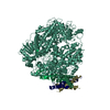
|
|---|---|
| 1 |
|
-Components
| #1: Protein | Mass: 233889.562 Da / Num. of mol.: 1 Source method: isolated from a genetically manipulated source Details: amino acid residues 1-25 is the N-terminal expression/purification tag containing a Strep II tag, a linker and a TEV protease cleavage site. Source: (gene. exp.) Human metapneumovirus (strain CAN97-83)Strain: CAN97-83 / Cell line (production host): Sf9 / Production host:   Spodoptera frugiperda (fall armyworm) / Strain (production host): IPLB-Sf21-AE Spodoptera frugiperda (fall armyworm) / Strain (production host): IPLB-Sf21-AEReferences: UniProt: Q6WB93, RNA-directed RNA polymerase, mRNA (guanine-N7)-methyltransferase, Transferases; Transferring phosphorus-containing groups; Nucleotidyltransferases, GDP polyribonucleotidyltransferase | ||
|---|---|---|---|
| #2: Protein | Mass: 35685.066 Da / Num. of mol.: 4 Source method: isolated from a genetically manipulated source Source: (gene. exp.) Human metapneumovirus (strain CAN97-83)Strain: CAN97-83 / Cell line (production host): Sf9 / Production host: Spodoptera frugiperda (fall armyworm) / Strain (production host): IPLB-Sf21-AE / References: UniProt: Q8B9Q8Has protein modification | N | |
-Experimental details
-Experiment
| Experiment | Method: ELECTRON MICROSCOPY |
|---|---|
| EM experiment | Aggregation state: PARTICLE / 3D reconstruction method: single particle reconstruction |
- Sample preparation
| Component | Name: Human metapneumovirus CAN97-83 / Type: COMPLEX Details: Human metapneumovirus L and P genes were co-expressed in sf9 insect cells using recombinant baculovirus expression system. The protein complex were affinity purified using nickel-NTA resin, ...Details: Human metapneumovirus L and P genes were co-expressed in sf9 insect cells using recombinant baculovirus expression system. The protein complex were affinity purified using nickel-NTA resin, followed by purification on a HiTrap Heparin column and a superpose 6 size exclusion column. Entity ID: all / Source: RECOMBINANT | |||||||||||||||||||||||||
|---|---|---|---|---|---|---|---|---|---|---|---|---|---|---|---|---|---|---|---|---|---|---|---|---|---|---|
| Molecular weight | Value: 0.36 MDa / Experimental value: YES | |||||||||||||||||||||||||
| Source (natural) | Organism: Human metapneumovirus CAN97-83 | |||||||||||||||||||||||||
| Source (recombinant) | Organism: Spodoptera frugiperda (fall armyworm) / Strain: IPLB-Sf21-AE / Cell: sf9 / Plasmid: pFastBacDual | |||||||||||||||||||||||||
| Natural host | Organism: homo sa | |||||||||||||||||||||||||
| Buffer solution | pH: 7.4 Details: sample of protein complex was in buffer containing 20 mM Na Hepes, pH 7.4, 300 mM NaCl, 1 mM MgCl2 and 0.5 mM TCEP (components as listed). | |||||||||||||||||||||||||
| Buffer component |
| |||||||||||||||||||||||||
| Specimen | Conc.: 0.8 mg/ml / Embedding applied: NO / Shadowing applied: NO / Staining applied: NO / Vitrification applied: YES Details: purified human metapneumovirus L:P complex in a 1:4 stoichiometry. | |||||||||||||||||||||||||
| Specimen support | Details: 20 mA / Grid material: COPPER / Grid mesh size: 200 divisions/in. / Grid type: Quantifoil R1.2/1.3 | |||||||||||||||||||||||||
| Vitrification | Instrument: FEI VITROBOT MARK IV / Cryogen name: ETHANE / Humidity: 100 % / Chamber temperature: 298 K Details: 3.5 uL sample (0.8 mg/mL)/grid, 4-second blotting (double-sided) at offset -2 before plunging. |
- Electron microscopy imaging
| Experimental equipment |  Model: Tecnai Polara / Image courtesy: FEI Company |
|---|---|
| Microscopy | Model: FEI POLARA 300 Details: preliminary grid screening was performed manually on a FEI Tecnai F20 microscope equipped with a Gatan K2 summit camera. |
| Electron gun | Electron source:  FIELD EMISSION GUN / Accelerating voltage: 300 kV / Illumination mode: OTHER FIELD EMISSION GUN / Accelerating voltage: 300 kV / Illumination mode: OTHER |
| Electron lens | Mode: BRIGHT FIELD / Nominal magnification: 31000 X / Calibrated magnification: 40323 X / Nominal defocus max: 3000 nm / Nominal defocus min: 1500 nm / Calibrated defocus min: 1500 nm / Calibrated defocus max: 3000 nm / Cs: 2.26 mm / C2 aperture diameter: 50 µm / Alignment procedure: COMA FREE |
| Specimen holder | Cryogen: NITROGEN / Specimen holder model: OTHER / Temperature (max): 90 K / Temperature (min): 77 K |
| Image recording | Average exposure time: 0.2 sec. / Electron dose: 1.2 e/Å2 / Detector mode: SUPER-RESOLUTION / Film or detector model: GATAN K2 SUMMIT (4k x 4k) / Num. of grids imaged: 1 / Num. of real images: 7438 |
| Image scans | Sampling size: 5 µm / Width: 7676 / Height: 7420 / Movie frames/image: 40 / Used frames/image: 1-40 |
- Processing
| Software | Name: PHENIX / Version: 1.16_3549: / Classification: refinement | ||||||||||||||||||||||||||||||||||||||||
|---|---|---|---|---|---|---|---|---|---|---|---|---|---|---|---|---|---|---|---|---|---|---|---|---|---|---|---|---|---|---|---|---|---|---|---|---|---|---|---|---|---|
| EM software |
| ||||||||||||||||||||||||||||||||||||||||
| CTF correction | Type: NONE | ||||||||||||||||||||||||||||||||||||||||
| Particle selection | Num. of particles selected: 5381943 | ||||||||||||||||||||||||||||||||||||||||
| Symmetry | Point symmetry: C1 (asymmetric) | ||||||||||||||||||||||||||||||||||||||||
| 3D reconstruction | Resolution: 3.7 Å / Resolution method: FSC 0.143 CUT-OFF / Num. of particles: 302346 / Symmetry type: POINT | ||||||||||||||||||||||||||||||||||||||||
| Refine LS restraints |
|
