Movie
Movie Controller


+ Open data

- Basic information
| Entry | Database: PDB / ID: 6tpq | ||||||
|---|---|---|---|---|---|---|---|
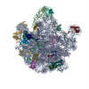
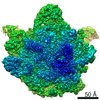
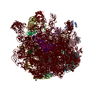
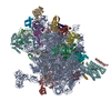
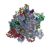
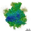
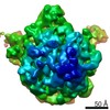
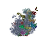
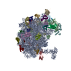
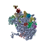
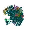
| Title | RNase M5 bound to 50S ribosome with precursor 5S rRNA | ||||||
 Components Components |
| ||||||
 Keywords Keywords | RIBOSOMAL PROTEIN / RNase M5 / M5 / complex / ribosome / 50S / RNA maturation / RNA processing / precursor 5S rRNA / pre-5S rRNA / 5S rRNA / L18 / Toprim domain / RNase | ||||||
| Function / homology |  Function and homology information Function and homology informationribonuclease M5 / ribonuclease M5 activity / positive regulation of rRNA processing / nucleoid / rRNA processing / large ribosomal subunit / transferase activity / 5S rRNA binding / ribosomal large subunit assembly / large ribosomal subunit rRNA binding ...ribonuclease M5 / ribonuclease M5 activity / positive regulation of rRNA processing / nucleoid / rRNA processing / large ribosomal subunit / transferase activity / 5S rRNA binding / ribosomal large subunit assembly / large ribosomal subunit rRNA binding / cytosolic large ribosomal subunit / cytoplasmic translation / tRNA binding / negative regulation of translation / rRNA binding / structural constituent of ribosome / ribosome / translation / ribonucleoprotein complex / mRNA binding / DNA binding / RNA binding / metal ion binding / cytoplasm Similarity search - Function | ||||||
| Biological species |   Geobacillus stearothermophilus (bacteria) Geobacillus stearothermophilus (bacteria) | ||||||
| Method | ELECTRON MICROSCOPY / single particle reconstruction / Resolution: 3.07 Å | ||||||
 Authors Authors | Oerum, S. / Dendooven, T. / Gilet, L. / Catala, M. / Degut, C. / Trinquier, A. / Barraud, P. / Luisi, B. / Condon, C. / Tisne, C. | ||||||
| Funding support |  France, 1items France, 1items
| ||||||
 Citation Citation | Journal: Mol Cell / Year: 2020 Title: Structures of B. subtilis Maturation RNases Captured on 50S Ribosome with Pre-rRNAs. Authors: Stephanie Oerum / Tom Dendooven / Marjorie Catala / Laetitia Gilet / Clément Dégut / Aude Trinquier / Maxime Bourguet / Pierre Barraud / Sarah Cianferani / Ben F Luisi / Ciarán Condon / Carine Tisné /  Abstract: The pathways for ribosomal RNA (rRNA) maturation diverge greatly among the domains of life. In the Gram-positive model bacterium, Bacillus subtilis, the final maturation steps of the two large ...The pathways for ribosomal RNA (rRNA) maturation diverge greatly among the domains of life. In the Gram-positive model bacterium, Bacillus subtilis, the final maturation steps of the two large ribosomal subunit (50S) rRNAs, 23S and 5S pre-rRNAs, are catalyzed by the double-strand specific ribonucleases (RNases) Mini-RNase III and RNase M5, respectively. Here we present a protocol that allowed us to solve the 3.0 and 3.1 Å resolution cryoelectron microscopy structures of these RNases poised to cleave their pre-rRNA substrates within the B. subtilis 50S particle. These data provide the first structural insights into rRNA maturation in bacteria by revealing how these RNases recognize and process double-stranded pre-rRNA. Our structures further uncover how specific ribosomal proteins act as chaperones to correctly fold the pre-rRNA substrates and, for Mini-III, anchor the RNase to the ribosome. These r-proteins thereby serve a quality-control function in the process from accurate ribosome assembly to rRNA processing. | ||||||
| History |
|
- Structure visualization
| Movie |
Movie viewer |
|---|---|
| Structure viewer | Molecule: MolmilJmol/JSmol |
- Downloads & links
-Download
| PDBx/mmCIF format | 6tpq.cif.gz | 2 MB | Display | PDBx/mmCIF format |
|---|---|---|---|---|
| PDB format | pdb6tpq.ent.gz | 1.6 MB | Display | PDB format |
| PDBx/mmJSON format | 6tpq.json.gz | Tree view | PDBx/mmJSON format | |
| Others |  Other downloads Other downloads |
-Validation report
| Arichive directory | https://data.pdbj.org/pub/pdb/validation_reports/tp/6tpqftp://data.pdbj.org/pub/pdb/validation_reports/tp/6tpq | HTTPS FTP |
|---|
-Related structure data
| Related structure data | 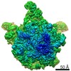 10543MC 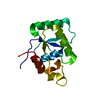 6tg6C 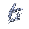 6tgjC  6tnnC M: map data used to model this data C: citing same article ( |
|---|---|
| Similar structure data |
-Links
 PDBj
PDBj


- Assembly
| Deposited unit | 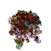
|
|---|---|
| 1 |
|
-Components
-Protein , 1 types, 2 molecules BA
| #1: Protein | Mass: 20778.699 Da / Num. of mol.: 2 Source method: isolated from a genetically manipulated source Source: (gene. exp.) Geobacillus stearothermophilus (bacteria)Gene: rnmV, B4109_0079, B4114_0027, D9548_05595, GT94_11730, TGS27_1231 Production host: |
|---|
-RNA chain , 2 types, 2 molecules VU
| #2: RNA chain | Mass: 39687.566 Da / Num. of mol.: 1 / Source method: isolated from a natural source Source: (natural) References: GenBank: 1150402534 |
|---|---|
| #4: RNA chain | Mass: 948320.438 Da / Num. of mol.: 1 / Source method: isolated from a natural source Source: (natural) References: GenBank: 467326 |
+50S ribosomal protein ... , 27 types, 27 molecules bWXYZacdefghijklmnopqrstuvw
-Non-polymers , 2 types, 222 molecules 

| #31: Chemical | ChemComp-MG /  Mass: 24.305 Da / Num. of mol.: 219 / Source method: obtained synthetically / Formula: Mg Mass: 24.305 Da / Num. of mol.: 219 / Source method: obtained synthetically / Formula: Mg#32: Chemical |  Mass: 65.409 Da / Num. of mol.: 3 / Source method: obtained synthetically / Formula: Zn Mass: 65.409 Da / Num. of mol.: 3 / Source method: obtained synthetically / Formula: Zn |
|---|
-Details
| Has ligand of interest | N |
|---|
-Experimental details
-Experiment
| Experiment | Method: ELECTRON MICROSCOPY |
|---|---|
| EM experiment | Aggregation state: PARTICLE / 3D reconstruction method: single particle reconstruction |
- Sample preparation
| Component |
| ||||||||||||||||||||||||
|---|---|---|---|---|---|---|---|---|---|---|---|---|---|---|---|---|---|---|---|---|---|---|---|---|---|
| Source (natural) |
| ||||||||||||||||||||||||
| Source (recombinant) | Organism: | ||||||||||||||||||||||||
| Buffer solution | pH: 7.5 | ||||||||||||||||||||||||
| Specimen | Embedding applied: NO / Shadowing applied: NO / Staining applied: NO / Vitrification applied: NO |
- Electron microscopy imaging
| Experimental equipment |  Model: Talos Arctica / Image courtesy: FEI Company |
|---|---|
| Microscopy | Model: FEI TALOS ARCTICA |
| Electron gun | Electron source:  FIELD EMISSION GUN / Accelerating voltage: 200 kV / Illumination mode: FLOOD BEAM FIELD EMISSION GUN / Accelerating voltage: 200 kV / Illumination mode: FLOOD BEAM |
| Electron lens | Mode: BRIGHT FIELD |
| Image recording | Electron dose: 23.94 e/Å2 / Film or detector model: FEI FALCON III (4k x 4k) |
- Processing
| EM software |
| ||||||||||||
|---|---|---|---|---|---|---|---|---|---|---|---|---|---|
| CTF correction | Type: PHASE FLIPPING AND AMPLITUDE CORRECTION | ||||||||||||
| 3D reconstruction | Resolution: 3.07 Å / Resolution method: FSC 0.143 CUT-OFF / Num. of particles: 92799 / Symmetry type: POINT | ||||||||||||
| Atomic model building | PDB-ID: 3J3V Accession code: 3J3V / Source name: PDB / Type: experimental model |