 Movie
Movie Controller

[English] 日本語
 Yorodumi
- PDB-6spf: Pseudomonas aeruginosa 70s ribosome from an aminoglycoside resist... -
+ Open data

- Basic information
| Entry | Database: PDB / ID: 6spf | |||||||||||||||
|---|---|---|---|---|---|---|---|---|---|---|---|---|---|---|---|---|
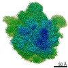
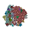
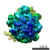
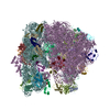
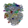
| Title | Pseudomonas aeruginosa 70s ribosome from an aminoglycoside resistant clinical isolate | |||||||||||||||
 Components Components |
| |||||||||||||||
 Keywords Keywords | RIBOSOME / Pseudomonas aeruginosa | |||||||||||||||
| Function / homology |  Function and homology information Function and homology informationlarge ribosomal subunit / transferase activity / ribosomal small subunit biogenesis / ribosomal small subunit assembly / small ribosomal subunit / 5S rRNA binding / ribosomal large subunit assembly / small ribosomal subunit rRNA binding / large ribosomal subunit rRNA binding / cytosolic small ribosomal subunit ...large ribosomal subunit / transferase activity / ribosomal small subunit biogenesis / ribosomal small subunit assembly / small ribosomal subunit / 5S rRNA binding / ribosomal large subunit assembly / small ribosomal subunit rRNA binding / large ribosomal subunit rRNA binding / cytosolic small ribosomal subunit / cytosolic large ribosomal subunit / cytoplasmic translation / tRNA binding / negative regulation of translation / rRNA binding / structural constituent of ribosome / ribosome / translation / ribonucleoprotein complex / mRNA binding / RNA binding / metal ion binding / cytoplasm / cytosol Similarity search - Function | |||||||||||||||
| Biological species |   Pseudomonas aeruginosa (bacteria) Pseudomonas aeruginosa (bacteria) | |||||||||||||||
| Method | ELECTRON MICROSCOPY / single particle reconstruction / cryo EM / Resolution: 2.89 Å | |||||||||||||||
 Authors Authors | Halfon, Y. / Jimenez-Fernande, A. / La Ros, R. / Espinos, R. / Krogh Johansen, H. / Matzov, D. / Eyal, Z. / Bashan, A. / Zimmerman, E. / Belousoff, M. ...Halfon, Y. / Jimenez-Fernande, A. / La Ros, R. / Espinos, R. / Krogh Johansen, H. / Matzov, D. / Eyal, Z. / Bashan, A. / Zimmerman, E. / Belousoff, M. / Molin, S. / Yonath, A. | |||||||||||||||
| Funding support |  Denmark, 4items Denmark, 4items
| |||||||||||||||
 Citation Citation | Journal: Proc Natl Acad Sci U S A / Year: 2019 Title: Structure of ribosomes from an aminoglycoside-resistant clinical isolate. Authors: Yehuda Halfon / Alicia Jimenez-Fernandez / Ruggero La Rosa / Rocio Espinosa Portero / Helle Krogh Johansen / Donna Matzov / Zohar Eyal / Anat Bashan / Ella Zimmerman / Matthew Belousoff / ...Authors: Yehuda Halfon / Alicia Jimenez-Fernandez / Ruggero La Rosa / Rocio Espinosa Portero / Helle Krogh Johansen / Donna Matzov / Zohar Eyal / Anat Bashan / Ella Zimmerman / Matthew Belousoff / Søren Molin / Ada Yonath /   Abstract: Resistance to antibiotics has become a major threat to modern medicine. The ribosome plays a fundamental role in cell vitality by the translation of the genetic code into proteins; hence, it is a ...Resistance to antibiotics has become a major threat to modern medicine. The ribosome plays a fundamental role in cell vitality by the translation of the genetic code into proteins; hence, it is a major target for clinically useful antibiotics. We report here the cryo-electron microscopy structures of the ribosome of a pathogenic aminoglycoside (AG)-resistant strain, as well as of a nonresistance strain isolated from a cystic fibrosis patient. The structural studies disclosed defective ribosome complex formation due to a conformational change of rRNA helix H69, an essential intersubunit bridge, and a secondary binding site of the AGs. In addition, a stable conformation of nucleotides A1486 and A1487, pointing into helix h44, is created compared to a non-AG-bound ribosome. We suggest that altering the conformations of ribosomal protein uL6 and rRNA helix H69, which interact with initiation-factor IF2, interferes with proper protein synthesis initiation. | |||||||||||||||
| History |
|
- Structure visualization
| Movie |
Movie viewer |
|---|---|
| Structure viewer | Molecule: MolmilJmol/JSmol |
- Downloads & links
-Download
| PDBx/mmCIF format | 6spf.cif.gz | 3 MB | Display | PDBx/mmCIF format |
|---|---|---|---|---|
| PDB format | pdb6spf.ent.gz | Display | PDB format | |
| PDBx/mmJSON format | 6spf.json.gz | Tree view | PDBx/mmJSON format | |
| Others |  Other downloads Other downloads |
-Validation report
| Summary document | 6spf_validation.pdf.gz | 1.2 MB | Display | wwPDB validaton report |
|---|---|---|---|---|
| Full document | 6spf_full_validation.pdf.gz | 1.4 MB | Display | |
| Data in XML | 6spf_validation.xml.gz | 200.5 KB | Display | |
| Data in CIF | 6spf_validation.cif.gz | 351.9 KB | Display | |
| Arichive directory | https://data.pdbj.org/pub/pdb/validation_reports/sp/6spfftp://data.pdbj.org/pub/pdb/validation_reports/sp/6spf | HTTPS FTP |
-Related structure data
| Related structure data | 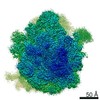 10284MC 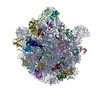 6spbC  6spcC 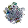 6spdC  6speC  6spgC M: map data used to model this data C: citing same article ( |
|---|---|
| Similar structure data |
-Links
 PDBj
PDBj


- Assembly
| Deposited unit | 
|
|---|---|
| 1 |
|
-Components
-RNA chain , 3 types, 3 molecules ABa
| #1: RNA chain | Mass: 935990.438 Da / Num. of mol.: 1 / Source method: isolated from a natural source / Source: (natural) Pseudomonas aeruginosa (bacteria) |
|---|---|
| #2: RNA chain | Mass: 37679.391 Da / Num. of mol.: 1 / Source method: isolated from a natural source / Source: (natural) Pseudomonas aeruginosa (bacteria) / References: GenBank: 1384417243 |
| #33: RNA chain | Mass: 493068.969 Da / Num. of mol.: 1 / Source method: isolated from a natural source / Source: (natural) Pseudomonas aeruginosa (bacteria) / References: GenBank: 1378074500 |
+50S ribosomal protein ... , 29 types, 29 molecules CDEFGHIJKLMNOPQRSTUVWXZ123456
-Protein , 1 types, 1 molecules Y
| #25: Protein | Mass: 6807.757 Da / Num. of mol.: 1 / Source method: isolated from a natural source / Source: (natural) Pseudomonas aeruginosa (bacteria) |
|---|
-30S ribosomal protein ... , 20 types, 20 molecules bcdefghijklmnopqrstu
| #34: Protein | Mass: 25366.418 Da / Num. of mol.: 1 / Source method: isolated from a natural source / Source: (natural) Pseudomonas aeruginosa (bacteria) / References: UniProt: A0A072ZPV8 |
|---|---|
| #35: Protein | Mass: 23180.906 Da / Num. of mol.: 1 / Source method: isolated from a natural source / Source: (natural) Pseudomonas aeruginosa (bacteria) / References: UniProt: A0A140S919 |
| #36: Protein | Mass: 23116.396 Da / Num. of mol.: 1 / Source method: isolated from a natural source / Source: (natural) Pseudomonas aeruginosa (bacteria) / References: UniProt: A0A072ZDF7 |
| #37: Protein | Mass: 15761.337 Da / Num. of mol.: 1 / Source method: isolated from a natural source / Source: (natural) Pseudomonas aeruginosa (bacteria) / References: UniProt: A0A241XG65 |
| #38: Protein | Mass: 11613.011 Da / Num. of mol.: 1 / Source method: isolated from a natural source / Source: (natural) Pseudomonas aeruginosa (bacteria) / References: UniProt: A0A069Q263 |
| #39: Protein | Mass: 17313.199 Da / Num. of mol.: 1 / Source method: isolated from a natural source / Source: (natural) Pseudomonas aeruginosa (bacteria) / References: UniProt: A0A2V3F2U6 |
| #40: Protein | Mass: 14001.312 Da / Num. of mol.: 1 / Source method: isolated from a natural source / Source: (natural) Pseudomonas aeruginosa (bacteria) / References: UniProt: E2RXT9 |
| #41: Protein | Mass: 14139.195 Da / Num. of mol.: 1 / Source method: isolated from a natural source / Source: (natural) Pseudomonas aeruginosa (bacteria) / References: UniProt: A0A069PXX1 |
| #42: Protein | Mass: 10886.569 Da / Num. of mol.: 1 / Source method: isolated from a natural source / Source: (natural) Pseudomonas aeruginosa (bacteria) / References: UniProt: E2RXT0 |
| #43: Protein | Mass: 12086.657 Da / Num. of mol.: 1 / Source method: isolated from a natural source / Source: (natural) Pseudomonas aeruginosa (bacteria) / References: UniProt: E2RXU4 |
| #44: Protein | Mass: 13472.612 Da / Num. of mol.: 1 / Source method: isolated from a natural source / Source: (natural) Pseudomonas aeruginosa (bacteria) / References: UniProt: A0A071L394 |
| #45: Protein | Mass: 12307.191 Da / Num. of mol.: 1 / Source method: isolated from a natural source / Source: (natural) Pseudomonas aeruginosa (bacteria) / References: UniProt: E2RXU3 |
| #46: Protein | Mass: 11203.024 Da / Num. of mol.: 1 / Source method: isolated from a natural source / Source: (natural) Pseudomonas aeruginosa (bacteria) / References: UniProt: E2RXT8 |
| #47: Protein | Mass: 9847.271 Da / Num. of mol.: 1 / Source method: isolated from a natural source / Source: (natural) Pseudomonas aeruginosa (bacteria) / References: UniProt: A0A071L3R7 |
| #48: Protein | Mass: 8762.938 Da / Num. of mol.: 1 / Source method: isolated from a natural source / Source: (natural) Pseudomonas aeruginosa (bacteria) / References: UniProt: A0A2V4FRZ2 |
| #49: Protein | Mass: 8848.358 Da / Num. of mol.: 1 / Source method: isolated from a natural source / Source: (natural) Pseudomonas aeruginosa (bacteria) / References: UniProt: E2RXT5 |
| #50: Protein | Mass: 6411.410 Da / Num. of mol.: 1 / Source method: isolated from a natural source / Source: (natural) Pseudomonas aeruginosa (bacteria) / References: UniProt: A0A2V3DLV3 |
| #51: Protein | Mass: 9182.882 Da / Num. of mol.: 1 / Source method: isolated from a natural source / Source: (natural) Pseudomonas aeruginosa (bacteria) / References: UniProt: E2RXT2 |
| #52: Protein | Mass: 9497.140 Da / Num. of mol.: 1 / Source method: isolated from a natural source / Source: (natural) Pseudomonas aeruginosa (bacteria) / References: UniProt: A0A072ZDZ9 |
| #53: Protein/peptide | Mass: 4209.891 Da / Num. of mol.: 1 / Source method: isolated from a natural source / Source: (natural) Pseudomonas aeruginosa (bacteria) / References: UniProt: A0A069QC99 |
-Details
| Has protein modification | Y |
|---|
-Experimental details
-Experiment
| Experiment | Method: ELECTRON MICROSCOPY |
|---|---|
| EM experiment | Aggregation state: PARTICLE / 3D reconstruction method: single particle reconstruction |
- Sample preparation
| Component | Name: Pseudomonas aeruginosa 70s ribosome from a clinical isolate Type: RIBOSOME / Entity ID: all / Source: NATURAL |
|---|---|
| Source (natural) | Organism: Pseudomonas aeruginos (bacteria) |
| Buffer solution | pH: 7.4 |
| Specimen | Embedding applied: NO / Shadowing applied: NO / Staining applied: NO / Vitrification applied: YES |
| Vitrification | Instrument: FEI VITROBOT MARK IV / Cryogen name: ETHANE / Humidity: 100 % / Chamber temperature: 277 K |
- Electron microscopy imaging
| Experimental equipment |  Model: Titan Krios / Image courtesy: FEI Company |
|---|---|
| Microscopy | Model: FEI TITAN KRIOS |
| Electron gun | Electron source:  FIELD EMISSION GUN / Accelerating voltage: 300 kV / Illumination mode: FLOOD BEAM FIELD EMISSION GUN / Accelerating voltage: 300 kV / Illumination mode: FLOOD BEAM |
| Electron lens | Mode: BRIGHT FIELD |
| Image recording | Electron dose: 1 e/Å2 / Film or detector model: FEI FALCON II (4k x 4k) |
- Processing
| EM software |
| |||||||||||||||||||||
|---|---|---|---|---|---|---|---|---|---|---|---|---|---|---|---|---|---|---|---|---|---|---|
| CTF correction | Type: NONE | |||||||||||||||||||||
| Symmetry | Point symmetry: C1 (asymmetric) | |||||||||||||||||||||
| 3D reconstruction | Resolution: 2.89 Å / Resolution method: FSC 0.143 CUT-OFF / Num. of particles: 319022 / Symmetry type: POINT |
