Movie
Movie Controller


+ Open data

- Basic information
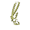
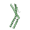
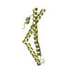
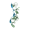
| Entry | Database: PDB / ID: 6rju | ||||||
|---|---|---|---|---|---|---|---|
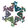
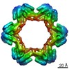
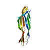
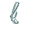
| Title | C-terminal domain of TssA protein from T6SS of Escherichia coli. | ||||||
 Components Components | Putative type VI secretion protein | ||||||
 Keywords Keywords | TRANSPORT PROTEIN / T6SS / E. coli / TssA / cap | ||||||
| Function / homology | Type VI secretion system-associated, VCA0119 / Type VI secretion, EvfE, EvfF, ImpA, BimE, VC_A0119, VasJ / ImpA, N-terminal / ImpA, N-terminal, type VI secretion system / Putative type VI secretion protein Function and homology information Function and homology information | ||||||
| Biological species |  | ||||||
| Method | ELECTRON MICROSCOPY / single particle reconstruction / cryo EM / Resolution: 3.2 Å | ||||||
 Authors Authors | Nazarov, S. / Shneider, M. / Basler, M. / Leiman, P. | ||||||
| Funding support |  Switzerland, 1items Switzerland, 1items
| ||||||
 Citation Citation | Journal: To Be Published Title: Cryo-EM structure of TssA protein from Type VI secretion system of E. coli. Authors: Nazarov, S. / Demurtas, D. / Shneider, M. / Basler, M. / Leiman, P. | ||||||
| History |
|
- Structure visualization
| Movie |
Movie viewer |
|---|---|
| Structure viewer | Molecule: MolmilJmol/JSmol |
- Downloads & links
-Download
| PDBx/mmCIF format | 6rju.cif.gz | 61.7 KB | Display | PDBx/mmCIF format |
|---|---|---|---|---|
| PDB format | pdb6rju.ent.gz | 40.5 KB | Display | PDB format |
| PDBx/mmJSON format | 6rju.json.gz | Tree view | PDBx/mmJSON format | |
| Others |  Other downloads Other downloads |
-Validation report
| Arichive directory | https://data.pdbj.org/pub/pdb/validation_reports/rj/6rjuftp://data.pdbj.org/pub/pdb/validation_reports/rj/6rju | HTTPS FTP |
|---|
-Related structure data
| Related structure data | 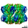 4906MC M: map data used to model this data C: citing same article ( |
|---|---|
| Similar structure data |


-Links
 PDBj
PDBj- Assembly
| Deposited unit | 
|
|---|---|
| 1 |
|
-Components
| #1: Protein | Mass: 59159.789 Da / Num. of mol.: 1 Source method: isolated from a genetically manipulated source Source: (gene. exp.) |
|---|---|
| Has protein modification | Y |
-Experimental details
-Experiment
| Experiment | Method: ELECTRON MICROSCOPY |
|---|---|
| EM experiment | Aggregation state: PARTICLE / 3D reconstruction method: single particle reconstruction |
- Sample preparation
| Component | Name: C-terminal domain of T6SS protein TssA. / Type: COMPLEX / Entity ID: all / Source: RECOMBINANT |
|---|---|
| Molecular weight | Value: 0.634 MDa / Experimental value: NO |
| Source (natural) | Organism: |
| Source (recombinant) | Organism: |
| Buffer solution | pH: 7.5 |
| Specimen | Embedding applied: NO / Shadowing applied: NO / Staining applied: NO / Vitrification applied: YES |
| Vitrification | Cryogen name: ETHANE / Humidity: 90 % / Chamber temperature: 293 K / Details: Leica EM GP2 |
- Electron microscopy imaging
| Experimental equipment |  Model: Titan Krios / Image courtesy: FEI Company |
|---|---|
| Microscopy | Model: FEI TITAN KRIOS |
| Electron gun | Electron source:  FIELD EMISSION GUN / Accelerating voltage: 300 kV / Illumination mode: FLOOD BEAM FIELD EMISSION GUN / Accelerating voltage: 300 kV / Illumination mode: FLOOD BEAM |
| Electron lens | Mode: BRIGHT FIELD / Cs: 2.7 mm / Alignment procedure: BASIC |
| Specimen holder | Cryogen: NITROGEN / Specimen holder model: FEI TITAN KRIOS AUTOGRID HOLDER |
| Image recording | Electron dose: 60 e/Å2 / Detector mode: COUNTING / Film or detector model: GATAN K2 SUMMIT (4k x 4k) / Num. of real images: 3269 |
| EM imaging optics | Energyfilter name: GIF Quantum LS |
| Image scans | Movie frames/image: 40 |
- Processing
| EM software |
| |||||||||||||||||||||||||||
|---|---|---|---|---|---|---|---|---|---|---|---|---|---|---|---|---|---|---|---|---|---|---|---|---|---|---|---|---|
| CTF correction | Type: NONE | |||||||||||||||||||||||||||
| Particle selection | Num. of particles selected: 667861 | |||||||||||||||||||||||||||
| Symmetry | Point symmetry: D6 (2x6 fold dihedral) | |||||||||||||||||||||||||||
| 3D reconstruction | Resolution: 3.2 Å / Resolution method: FSC 0.143 CUT-OFF / Num. of particles: 52370 / Symmetry type: POINT | |||||||||||||||||||||||||||
| Atomic model building | Protocol: RIGID BODY FIT | |||||||||||||||||||||||||||
| Atomic model building | PDB-ID: 4YO5 Pdb chain-ID: A / Accession code: 4YO5 / Pdb chain residue range: 388-474 / Source name: PDB / Type: experimental model |